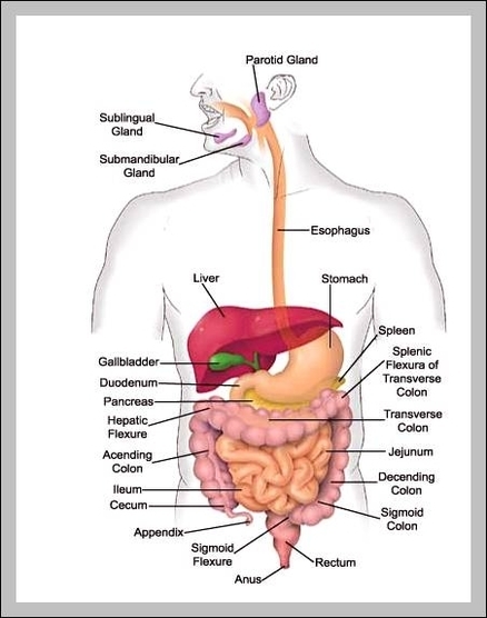
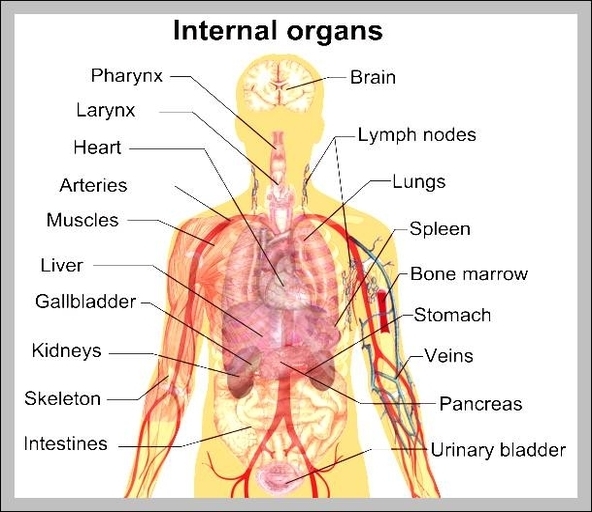
Small Intestine Anatomy
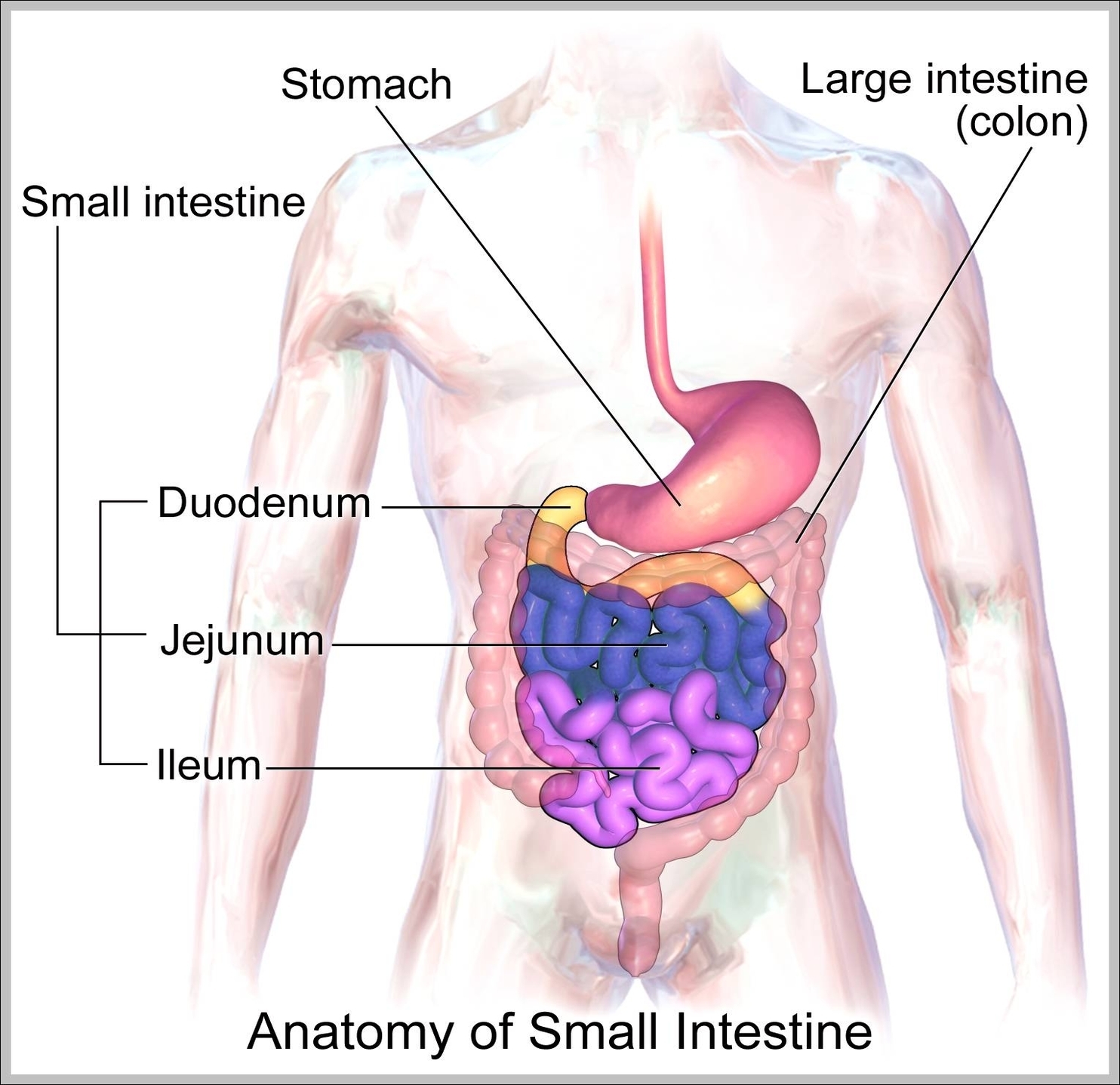
Anatomy The small intestine is made up of thee sections, including the duodenum, the jejunum and the ileum. On its proximal (near) end, the small intestine—beginning with the duodenum—connects to the stomach. On its distal (far) end, the ileum—the last View Diagram Small Intestine Anatomy