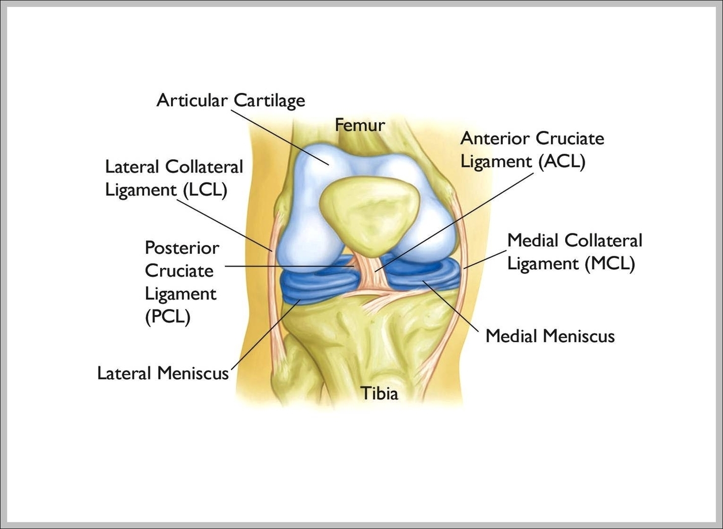
Ankle Ligaments Image
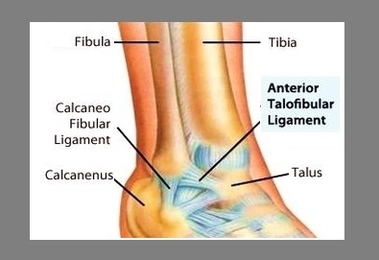
There are several major ligaments in the ankle: Three ligaments on the outside of the ankle that make up the lateral ligament complex, as follows: The anterior talofibular ligament (ATFL), which connects the front of the talus bone to the View Diagram Ankle Ligaments Image