Thymus Diagram Image

This human anatomy diagram with labels depicts and explains the details and or parts of the Thymus Diagram Image. Human anatomy diagrams and charts show internal organs, body systems, cells, conditions, sickness and symptoms information and/or tips to ensure one lives in good health.

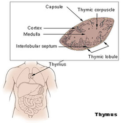
The gland extends from as high as the lower border of the thyroid gland to the fourth costal cartilage downwards. The thymus is of a pinkish-grey color, soft, and lobulated on its surfaces. Embryologically it is derived from the third pharyngeal pouch . The thymus is the first of the lymphoid organs to be formed.

The thymus gland is a soft bilobed organ which is encapsulated. It lies in the superior mediastinum and in the anterior part of the inferior mediastinum, close to the pericardium, anterior to the great vessels of the heart, and deep to the sternum. It extends from the level of the inferior poles thyroid gland above, to the fourth costal cartilage.

The thymus is a primary lymphoid organ located in the mediastinum. It consists of two lobes connected by an isthmus. Histologically, the thymus is divided into lobules, each one consisting of a central medulla and a peripheral cortex.

Thymus Diagram Image

Thymus Diagram Image


Tags: