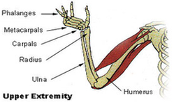
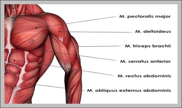
Upper Extremity Diagram1 Image

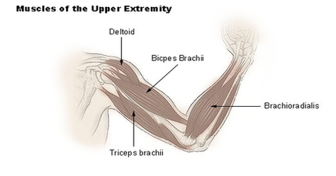
The forearm is the portion between the elbow and wrist. The thigh is the portion of the lower extremity between the hip and knee, and the calf is the portion between the knee and ankle. The normal arterial anatomy of View Diagram Upper Extremity Diagram1 Image