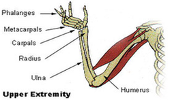
Anatomy of the upper extremity in standard radiology This radioanatomy module on the upper limb presents 16 radiographic images with 112 structures labeled.
The forearm is the portion between the elbow and wrist. The thigh is the portion of the lower extremity between the hip and knee, and the calf is the portion between the knee and ankle. The normal arterial anatomy of the upper extremity is depicted graphically in Figure 13-1.
Plain X-Rays of the upper limb The first X-Ray image is focused on the pectoral girdle seen from the front, allowing us to study the clavicles, sternum and sternoclavicular joints. The following image shows the clavicle in an AP view, with its sternal and acromial extremities, conoid tubercle and the body of the clavicle.
Upper Extremity Diagram Image