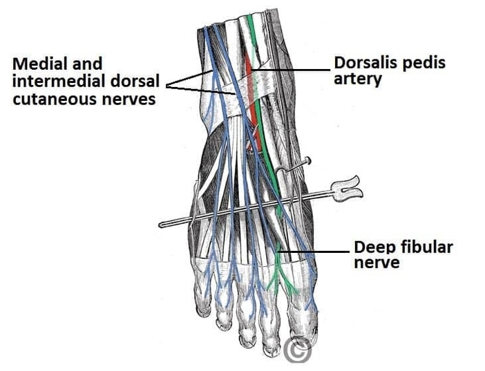
The nerves of the dorsum of the foot provide sensory innervation to the skin of the dorsal surface. This anatomy diagram of the anatomy of the nerves of the dorsum of the foot highlights the superficial and deep fibular nerves. It shows how the superficial fibular nerve supplies most of the dorsum, while the deep fibular nerve innervates the first web space. The diagram clarifies their relationship to extensor tendons and ankle structures. Understanding this distribution is essential in assessing nerve injuries and foot trauma. This visual guide supports lower limb neuroanatomy learning. Anatomy of the Nerves of the Dorsum of the Foot Superficial and Deep Fibular Nerves Diagram - Chart - diagrams and charts with labels. This diagram depicts Anatomy of the Nerves of the Dorsum of the Foot Superficial and Deep Fibular Nerves and explains the details of Anatomy of the Nerves of the Dorsum of the Foot Superficial and Deep Fibular Nerves.
Anatomy of the Nerves of the Dorsum of the Foot Superficial and Deep Fibular Nerves