Terminal Ileum Anatomy 2 Image

This human anatomy diagram with labels depicts and explains the details and or parts of the Terminal Ileum Anatomy 2 Image. Human anatomy diagrams and charts show internal organs, body systems, cells, conditions, sickness and symptoms information and/or tips to ensure one lives in good health.

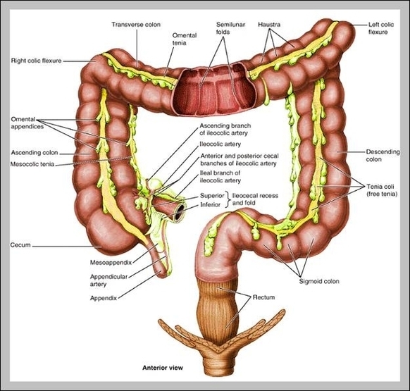
Terminal Ileum. The terminal ileum is the distal end of the small intestine that intersects with the large intestine.

Ileum (“twisted intestine”) is the final and the longest portion of the small intestine, the ileum measures about 3 m (9.8 ft ) and contributes to almost 60% of the length of the small intestine. The ileum joins the large intestine at a smooth muscle sphincter called the ileocecal sphincter (valve).

Characteristics & Features. The transition from the jejunum to the ileum is not sharply marked, while at the distal end, the ileum opens into the large intestine. At the junction between the ileum and the caecum lies the ileocecal valve (ileal ostium), a functional sphincter formed by the circular muscle layers of both the ileum and cecum.

Terminal Ileum Anatomy 2 Image

Terminal Ileum Anatomy 2 Image


Tags: , ,