Skip to content

Human Anatomy Diagrams

Menu

Primary menu

  • Top
  • Anatomy
  • Muscles
  • Systems
  • Body Parts
  • Organs
  • Cells
  • Bones
  • Teeth
  • Cancer
  • Health

Category Archives: System

Human anatomy systems diagrams and charts

Endocrine System

Posted on October 18, 2025 by anatomy
Endocrine System

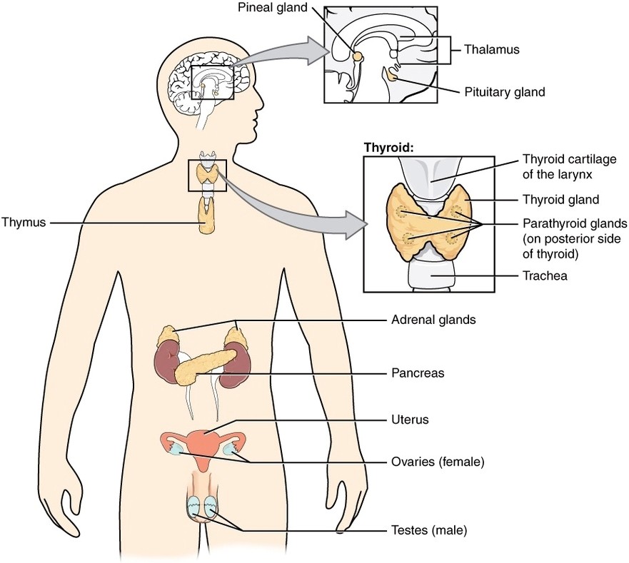
Endocrine System: The endocrine system is a network of glands that release hormones into the bloodstream to regulate processes like metabolism, growth, reproduction, and homeostasis.

Posted in System | Tagged adrenal glands, endocrine system diagram, glands function, hormone regulation, thyroid

Baroreceptor Reflex Flow Art

Posted on October 1, 2025 by anatomy
Baroreceptor Reflex Flow Art

Baroreceptor Reflex Flow Art: The baroreceptor reflex helps maintain blood pressure homeostasis by detecting changes in arterial stretch and triggering heart rate and vessel tone adjustments via the autonomic nervous system.

Posted in System | Tagged autonomic control, baroreceptor diagram, baroreflex pathway, blood pressure regulation, cardiovascular reflex

The Hematopoietic System of the Bone Marrow new

Posted on September 30, 2025 by anatomy
The Hematopoietic System of the Bone Marrow new

The Hematopoietic System of the Bone Marrow new: The bone marrow’s hematopoietic system consists of stem cells that differentiate into various blood cell types, providing a continuous supply of cells vital for immunity, oxygen transport, and clotting.

Posted in System | Tagged blood cell formation, bone marrow diagram, hematopoiesis process, immune system origin, stem cells

Renin Aldosterone Angiotensin

Posted on September 29, 2025 by anatomy
Renin Aldosterone Angiotensin

Renin Aldosterone Angiotensin: The renin-angiotensin-aldosterone system (RAAS) regulates blood pressure and fluid balance by promoting vasoconstriction and sodium retention in response to decreased blood volume or pressure.

Posted in System | Tagged blood pressure regulation, fluid balance, hormone signaling, kidney function, RAAS pathway diagram

Cardiac Cycle vs Heart Sounds

Posted on September 28, 2025 by anatomy
Cardiac Cycle vs Heart Sounds

Cardiac Cycle vs Heart Sounds: The cardiac cycle corresponds with heart sounds, where the “lub” (S1) is caused by AV valve closure during systole, and the “dub” (S2) marks semilunar valve closure during diastole.

Posted in System | Tagged cardiac cycle and heart sound diagram, ECG comparison, heart contraction sounds, lub dub explanation, valve activity

Automatic Innervation

Posted on September 4, 2025 by anatomy
Automatic Innervation

Automatic Innervation: Autonomic innervation involves the sympathetic and parasympathetic nervous systems, which regulate involuntary functions like heart rate, digestion, and respiratory rate through motor neurons.

Posted in System | Tagged automatic control, autonomic nervous system diagram, involuntary functions, nervous system control, sympathetic and parasympathetic

Dual System of Human Circulation

Posted on September 1, 2025 by anatomy
Dual System of Human Circulation

Dual System of Human Circulation: The human circulatory system has two circuits: the pulmonary circuit carries deoxygenated blood to the lungs, while the systemic circuit distributes oxygenated blood to the body.

Posted in System | Tagged cardiovascular system, circulatory pathways, dual circulation system diagram, heart circulation, systemic and pulmonary circuits

Cardiac Cycle vs Electrocardiogram

Posted on August 26, 2025 by anatomy
Cardiac Cycle vs Electrocardiogram

Cardiac Cycle vs Electrocardiogram: The cardiac cycle represents mechanical heart activity, while the electrocardiogram shows its electrical activity; both correlate to diagnose rhythm and timing issues.

Posted in System | Tagged cardiac cycle diagram, ECG vs heart cycle, electrocardiogram interpretation, heart rhythm, heartbeats

The Enzyme Renin Converts The Pro Enzyme Angiotensin I

Posted on August 20, 2025 by anatomy
The Enzyme Renin Converts The Pro Enzyme Angiotensin I

The Enzyme Renin Converts The Pro Enzyme Angiotensin I: Renin, secreted by the kidneys, catalyzes the conversion of angiotensinogen to angiotensin I, which is further converted to angiotensin II, a vasoconstrictor that raises blood pressure.

Posted in System | Tagged blood pressure regulation, enzyme function, hormone pathway, renin conversion, renin-angiotensin system diagram

The Hypothalamus Controls Thermoregulation

Posted on August 15, 2025 by anatomy
The Hypothalamus Controls Thermoregulation

The Hypothalamus Controls Thermoregulation: The hypothalamus monitors body temperature and triggers responses like sweating or shivering to maintain homeostasis through its control of the autonomic nervous system.

Posted in System | Tagged body heat control, endocrine function, hypothalamus thermoregulation diagram, temperature regulation, thermoregulatory center

Net Filtration Pressure

Posted on August 8, 2025 by anatomy
Net Filtration Pressure

Net Filtration Pressure: Net filtration pressure (NFP) is the balance of hydrostatic and osmotic pressures in the glomerulus, determining the movement of fluid into the nephron for urine formation.

Posted in System | Tagged fluid movement, glomerular filtration, kidney function, net filtration pressure diagram, renal filtration

Conduction System of Heart

Posted on August 6, 2025 by anatomy
Conduction System of Heart

Conduction System of Heart: The heart’s conduction system includes the SA node, AV node, bundle of His, bundle branches, and Purkinje fibers, coordinating the electrical impulses that drive rhythmic contractions.

Posted in System | Tagged AV node, electrical impulses, heart conduction system diagram, heart rhythm, SA node

Factors in Cardiac Output

Posted on July 22, 2025 by anatomy
Factors in Cardiac Output

Factors in Cardiac Output: Cardiac output depends on stroke volume and heart rate, influenced by preload, afterload, contractility, and autonomic nervous system regulation.

Posted in System | Tagged blood flow regulation, cardiac output diagram, cardiac performance, circulatory physiology, heart rate and stroke volume

Cardiac Conduction

Posted on July 13, 2025 by anatomy
Cardiac Conduction

Cardiac Conduction: The heart’s electrical system begins with the sinoatrial (SA) node, spreading impulses through the atria, to the atrioventricular (AV) node, and down the bundle branches and Purkinje fibers to coordinate contraction.

Posted in System | Tagged AV node, cardiac conduction system diagram, heart electrical pathway, Purkinje fibers, SA node

Neonatal Circulatory System

Posted on June 29, 2025 by anatomy
Neonatal Circulatory System

Neonatal Circulatory System: At birth, the neonatal circulatory system undergoes major changes as fetal shunts close and the lungs take over oxygen exchange, resulting in the adult-like pattern of blood flow.

Posted in System | Tagged ductus arteriosus, fetal to neonatal transition, foramen ovale closure, infant heart, newborn circulation diagram

Carbon Digestion

Posted on June 20, 2025 by anatomy
Carbon Digestion

Carbon Digestion: Carbohydrate digestion starts in the mouth with salivary amylase and continues in the small intestine with pancreatic enzymes, breaking polysaccharides into monosaccharides like glucose for absorption.

Posted in System | Tagged carbohydrate metabolism, carbon digestion diagram, digestive enzymes, energy production, glucose breakdown

Action Potential Heart Contraction

Posted on June 4, 2025 by anatomy
Action Potential Heart Contraction

Action Potential Heart Contraction: A cardiac action potential initiates when the SA node fires, spreading electrical impulses that trigger calcium influx, leading to coordinated contraction of the atria and ventricles.

Posted in System | Tagged action potential diagram heart, cardiac physiology, ECG interpretation, heart electrical signal, myocardial contraction

Blood Flow Relaxed Ventricles

Posted on May 25, 2025 by anatomy
Blood Flow Relaxed Ventricles

Blood Flow Relaxed Ventricles: When the ventricles relax during diastole, blood flows passively from the atria into the ventricles through the open AV valves, preparing the heart for the next contraction.

Posted in System | Tagged blood circulation, blood flow diagram, cardiac physiology, heart cycle, ventricular relaxation

Phases of the Cardiac Cycle

Posted on May 19, 2025 by anatomy
Phases of the Cardiac Cycle

Phases of the Cardiac Cycle: The cardiac cycle includes atrial systole, ventricular systole, and diastole, each phase coordinating contraction and relaxation to maintain continuous blood flow through the heart and circulatory system.

Posted in System | Tagged cardiac cycle diagram, ECG interpretation, heart contraction phases, heart rhythm, systole and diastole

Depolarization in Sinus Rhythm

Posted on May 11, 2025 by anatomy
Depolarization in Sinus Rhythm

Depolarization in Sinus Rhythm: In normal sinus rhythm, depolarization originates in the SA node and spreads uniformly through the atria and ventricles, resulting in coordinated heartbeats seen as distinct P, QRS, and T waves on an ECG.

Posted in System | Tagged cardiac electrophysiology, ECG waveform, heart depolarization, PQRST complex, sinus rhythm diagram

Post navigation

← Older posts
Newer posts →

Primary Sidebar Widget Area

Top Diagrams

  • Human Body Organ Locations Human Body Organ Locations

  • Map Of Body Muscles Map Of Body Muscles

  • Human Organs Back View Human Organs Back View

  • Female Abdominal Anatomy Female Abdominal Anatomy

  • Spinal Vertebrae Chart Spinal Vertebrae Chart

  • Map Of Human Organs Map Of Human Organs

  • Male Body Organs Diagram Male Body Organs Diagram

  • Female Muscular System Female Muscular System

  • Picture Of Organs On Left Side Of Body Picture Of Organs On Left Side Of Body

  • Lower Back Hip Anatomy Lower Back Hip Anatomy

  • Male Anatomy Pictures Male Anatomy Pictures

  • Female Human Anatomy Organs Female Human Anatomy Organs

  • Lower Back Anatomy Pictures Lower Back Anatomy Pictures

  • Muscular System Full Body Diagram Muscular System Full Body Diagram

  • Groin Muscles Anatomy Groin Muscles Anatomy

Latest Posts

  • Borders of the Femoral Triangle
  • Palmar Fascia of the Hand
  • Knee 3D
  • Anatomical Position of the Lungs
  • X Ray of Shoulder Separation
  • Aberrant and Supernumerary Arteries of the Kidney
  • Anatomical Location of the Lacrimal Gland
  • Superficial Cerebral Veins
  • Overview of the Anatomical Position of the Ulna
  • Human Skeletal System Diagram
  • Glomerulus
  • Posterior Surface of the Distal Portion of the Femur
  • Layers of the Oesophagus
  • Anatomy of the Fibula
  • Submandibular Triangle of the Neck Borders

Categories

  • Anatomy
  • Body Parts
  • Bones
  • Cancer
  • Cells
  • Health
  • Muscles
  • Organs
  • System
  • Teeth
  • Top

Top Tags

  • muscles (205)
  • muscle (171)
  • human (156)
  • human muscles (138)
  • muscles diagram (138)
  • muscles anatomy (138)
  • human muscle diagram (136)
  • muscular system (136)
  • body (119)
  • system (119)
  • body organs (119)
  • blood (82)
  • parts of a human body (77)
  • human body parts (77)
  • body parts (77)
  • organs (67)
  • organ (67)
  • pregnancy (66)
  • diabetes (62)
  • organ anatomy (62)
Copyright © 2026 . All Rights Reserved.
Theme: Catch Box by Catch Themes
Scroll Up