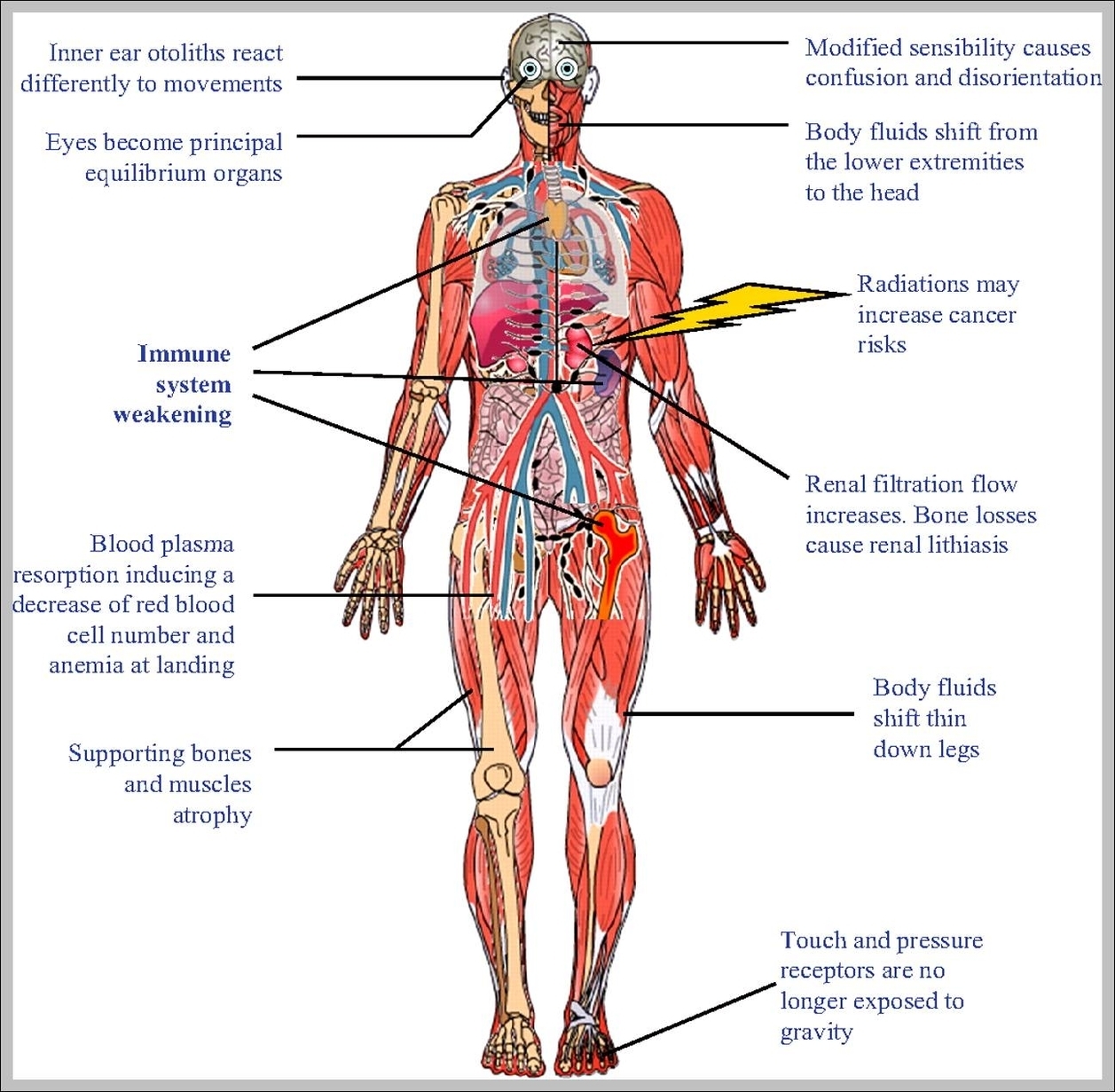
Download and print these Digestive System coloring pages for free. Digestive System coloring pages are a fun way for kids of all ages to develop creativity, focus, motor skills and color recognition.
Your digestive (say: dye-JES-tiv) system started working even before you took the first bite of your pizza. And the digestive system will be busy at work on your chewed-up lunch for the next few hours — or sometimes days, depending upon what you’ve eaten.
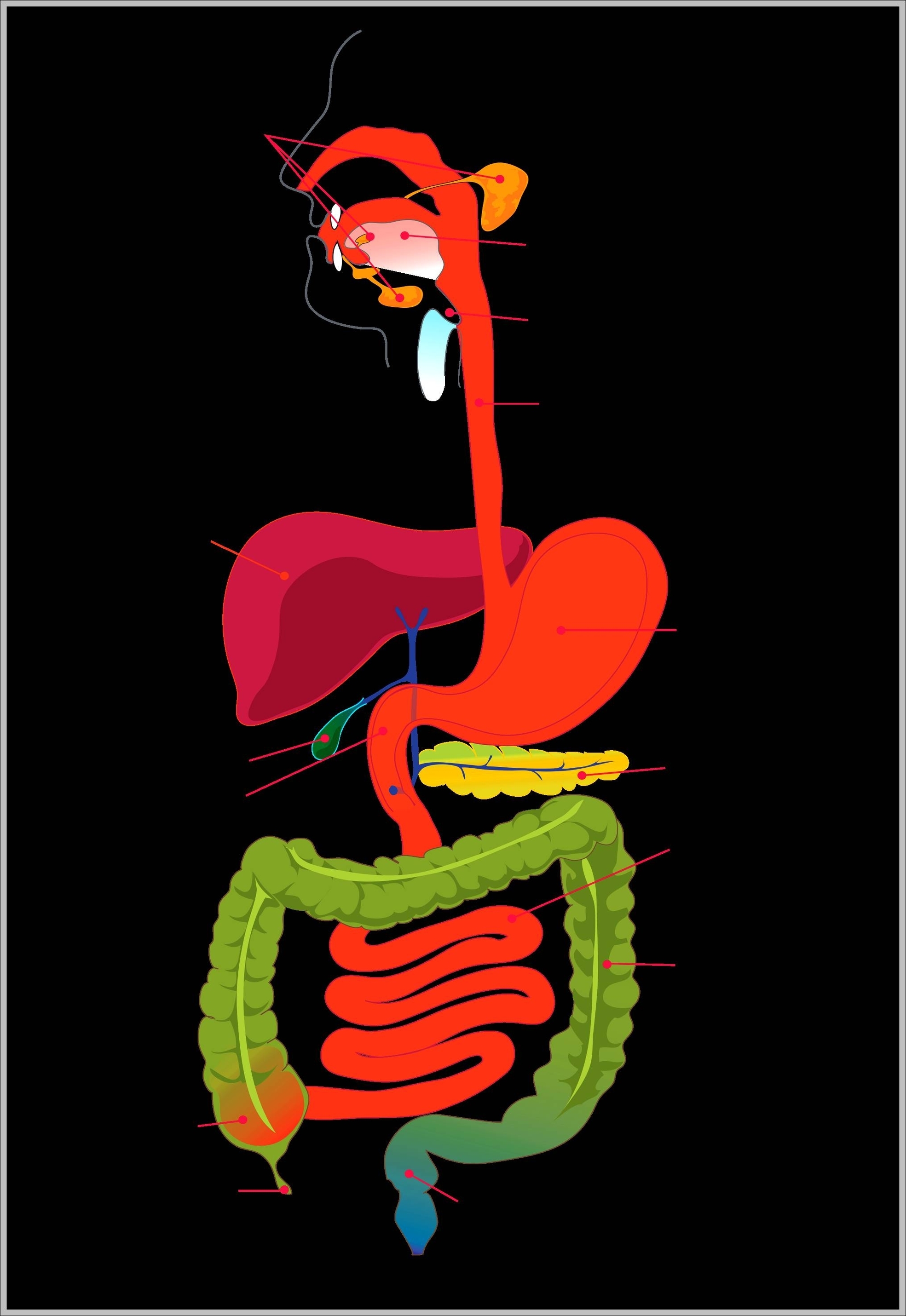
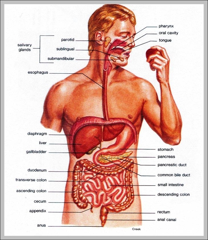
The mouth is where the digestive tract begins. Enzymes released into the mouth start the process of digestion. The epiglottis is a small piece of tissue that covers the opening of the larynx.
Pictures Of Digestive System For Kids Diagram - Chart - diagrams and charts with labels. This diagram depicts Pictures Of Digestive System For Kids