Sliding Filament Model Muscle Ultrastructure
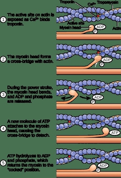
The sliding filament model explains how muscle contraction occurs at the microscopic level. Within each sarcomere, thin actin filaments slide past thick myosin filaments. When stimulated, myosin heads bind to actin and pull them inward, shortening the sarcomere. This process View Diagram Sliding Filament Model Muscle Ultrastructure