Colon Cancer Colon Diagram Image
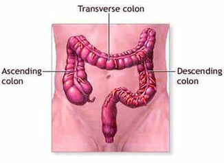
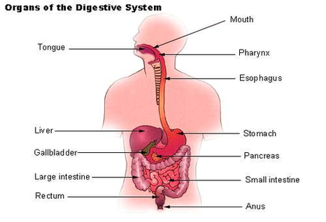
Colorectal cancer is cancer that occurs in the colon or rectum. Sometimes it is called colon for short. As the drawing shows, the colon is the large intestine or large bowel. 4,035 colorectal cancer stock photos and images available, or View Diagram Colon Cancer Colon Diagram Image