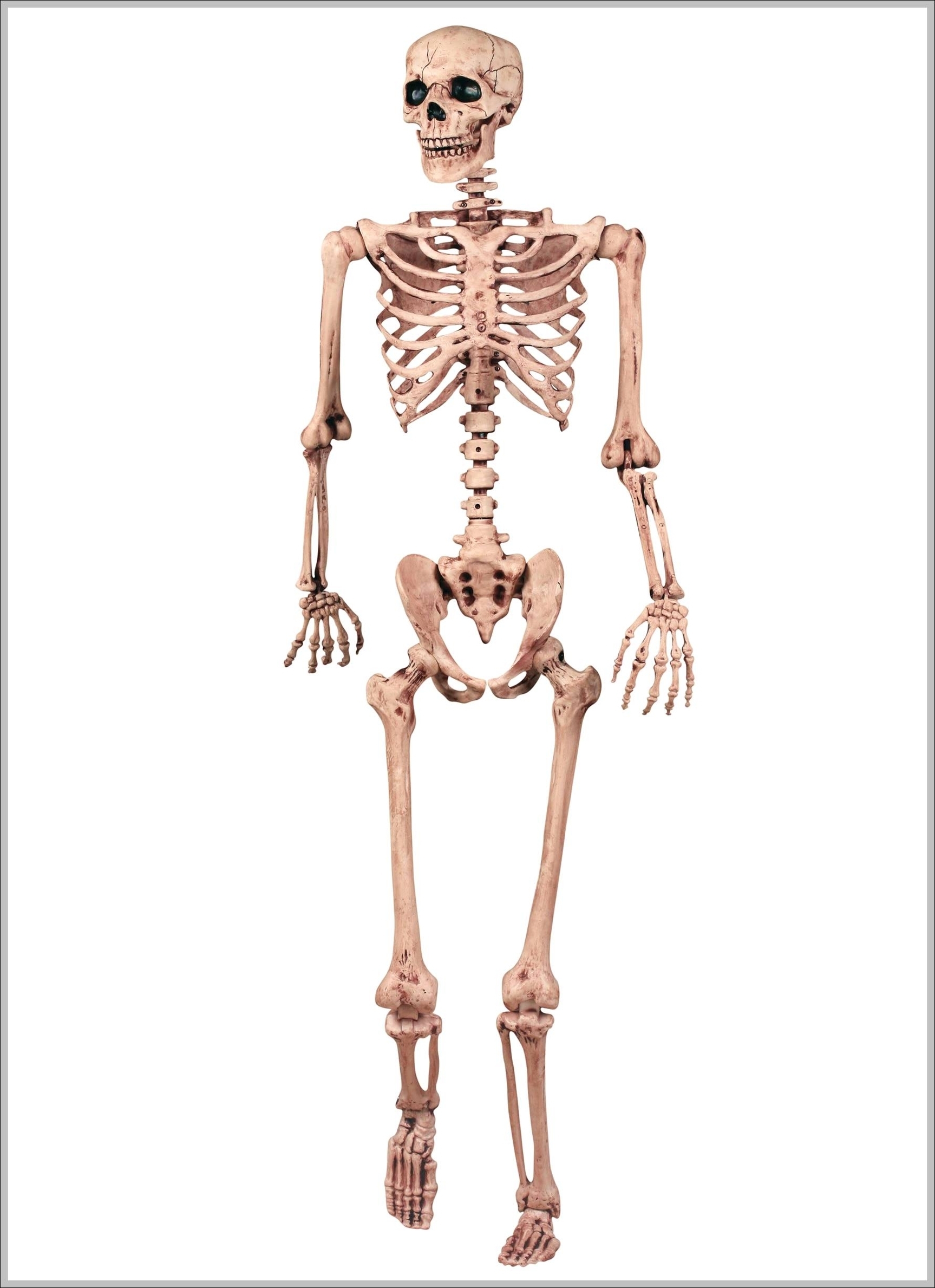
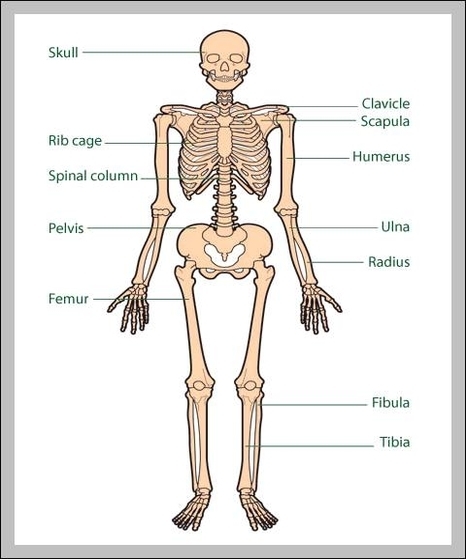
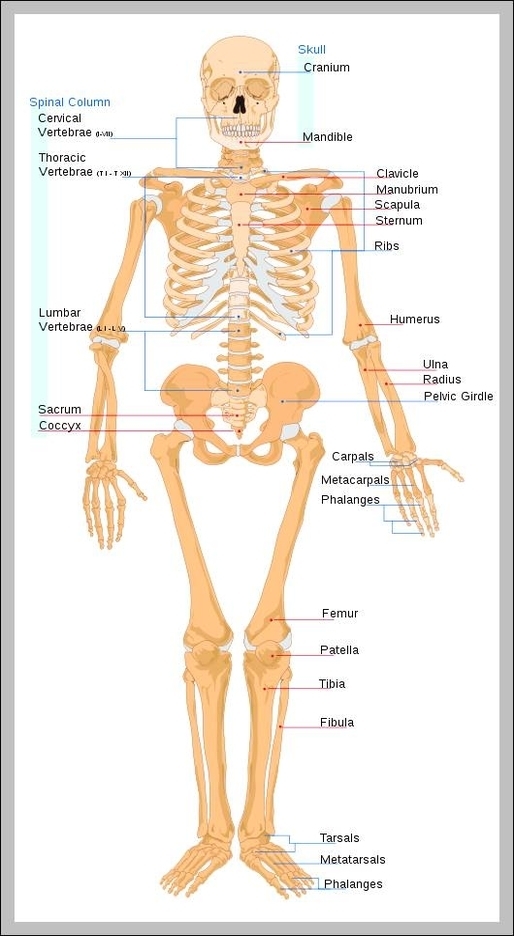
What Are The Functions Of The Skeletal System
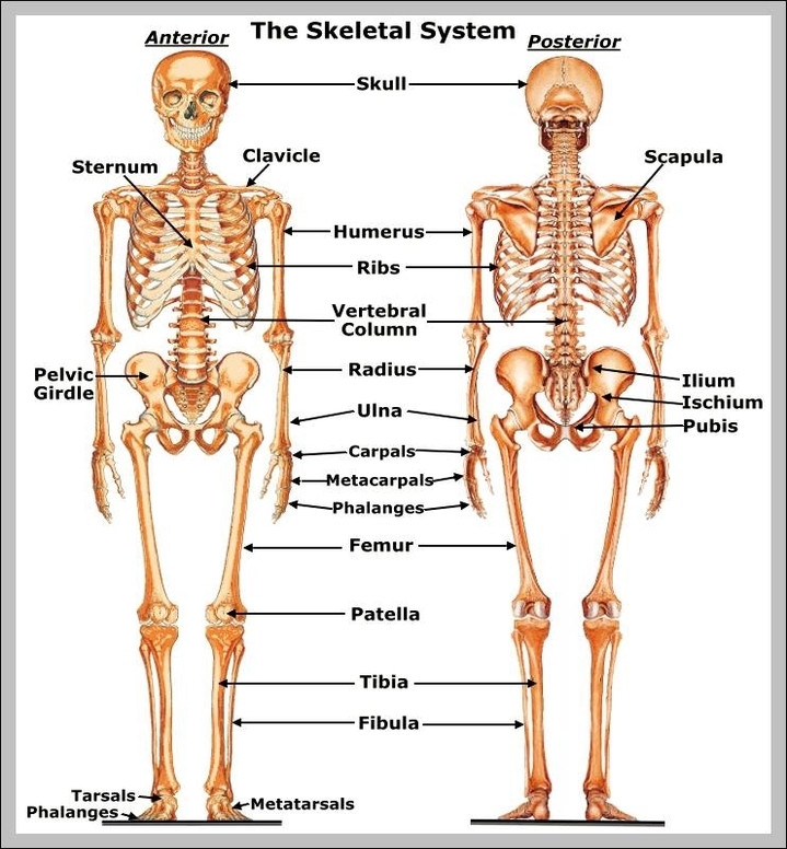
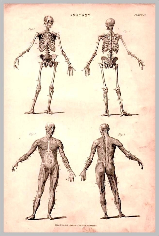
The skeletal system is the body system composed of bones and cartilage and performs the following critical functions for the human body: supports the body. facilitates movement. protects internal organs. produces blood cells. stores and releases minerals and fat. The View Diagram What Are The Functions Of The Skeletal System