Hand Anatomy Nerves Image

This human anatomy diagram with labels depicts and explains the details and or parts of the Hand Anatomy Nerves Image. Human anatomy diagrams and charts show internal organs, body systems, cells, conditions, sickness and symptoms information and/or tips to ensure one lives in good health.

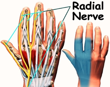
7,371 hand nerve anatomy stock photos, vectors, and illustrations are available royalty-free. See hand nerve anatomy stock video clips

The nerve powers almost all of the small muscles in the hand including the hypothenar muscles, the lumbricals to the ring and small finger, the palmar and dorsal interossei muscles, the adductor pollicus, and the deep head of the flexor pollicus brevis.

Anatomy of the Hand and Wrist: Bones, Muscles, Tendons, Nerves. The wrist links the hand to the arm. The wrist is a complex mechanical system of 8 small bones known as the carpal bones. The carpal bones are arranged in 2 interrelated rows. One row connects with the ends of the bones in the forearm— radius and ulna.

Hand Anatomy Nerves Image

Hand Anatomy Nerves Image


Tags: ,