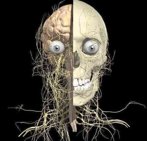
Head Nerves Anatomy Image

This human anatomy diagram with labels depicts and explains the details and or parts of the Head Nerves Anatomy Image. Human anatomy diagrams and charts show internal organs, body systems, cells, conditions, sickness and symptoms information and/or tips to ensure one lives in good health.

Nerves of the Head and Neck. The nerves of the head and neck include the most vital and important organs of the nervous system — the brain and spinal cord — as well as the organs of the special senses. In addition, in this region we also find the major cranial and spinal nerves that connect the central nervous system to the skin,…

Toggle Anatomy System. The muscles of the head and neck are also controlled by various cranial nerves including the facial nerve (facial expression) and accessory nerve (head and neck movements). Wandering through the neck and torso, the vagus nerve communicates vital information from the brain to the heart and intestines.

Each nerve also has a descriptive name (e.g. olfactory, optic, etc.) that identifies its function or location. The cranial nerves provide a direct connection to the brain for the special sense muscles of the head, neck, and shoulders, the heart, and the GI tract. Spinal Nerves.

Head Nerves Anatomy Image

Head Nerves Anatomy Image


Tags: ,