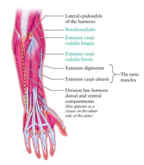
Muscles Just like the arm, the forearm is divided into two compartments by deep fascia; the interosseous membrane, and the fibrous intermuscular septa. This creates an anterior compartment that contains the flexor muscles, and a posterior one that contains the extensor muscles. Extensors of the forearm
Extending from the wrist to the elbow joint is the region of the upper extremity called the forearm (antebrachium). The forearm helps the shoulder and the arm in force application and the precise placement of the hand in space, with the help of the elbow and radioulnar joints.
– Flexors: superficial (flexor carpi ulnaris, palmaris longus, flexor carpi radialis, and pronator teres), intermediate (flexor digitorum superficialis, flexor digitorum profundus, and flexor pollicis longus) and deep (pronator quadratus). The forearm consists of two long bones; the radius and the ulna.
Diagram Dorsal Forearm Diagflat Image

