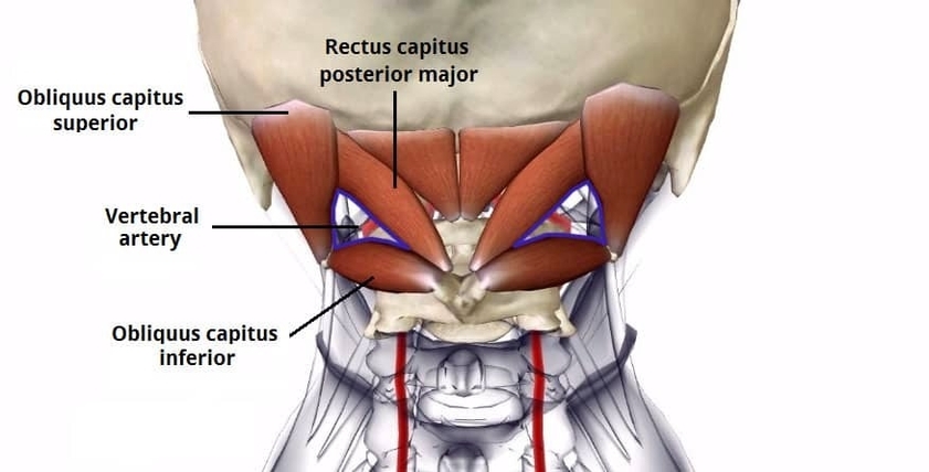
The suboccipital triangle, located deep in the posterior neck, is bordered by the rectus capitis posterior major, obliquus capitis superior, and obliquus capitis inferior muscles. The vertebral artery and suboccipital nerve pass through this space. Knowledge of its borders and contents is essential in neurology, cervical spine surgery, and imaging interpretation. Understanding its anatomy helps prevent injury to the vertebral artery during surgical procedures, guide nerve blocks, and localize lesions affecting neck or posterior cranial circulation. Precise anatomical insight ensures safe interventions while preserving vascular and neural integrity in this region. Suboccipital Triangle Borders and Vertebral Artery Diagram - Chart - diagrams and charts with labels. This diagram depicts Suboccipital Triangle Borders and Vertebral Artery and explains the details of Suboccipital Triangle Borders and Vertebral Artery.
Suboccipital Triangle Borders and Vertebral Artery