Scan Plantar Foot Image

This human anatomy diagram with labels depicts and explains the details and or parts of the Scan Plantar Foot Image. Human anatomy diagrams and charts show internal organs, body systems, cells, conditions, sickness and symptoms information and/or tips to ensure one lives in good health.

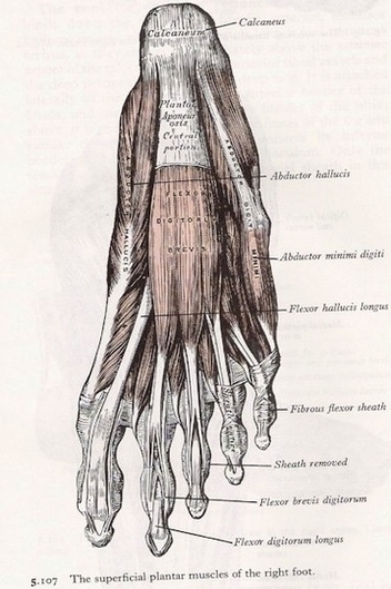
A diagnostic ultrasound, also called a sonogram, is a quick and easy imaging method used to take a look at soft tissues in the foot (not to be confused with Ultrasound Therapy for treating Plantar Fasciitis). This imaging tool can rule out soft-tissue conditions like tendonitis, tarsal tunnel, nerve compression, and plantar fibromatosis.

The plantar aspect of the foot is seen at the top of the image. (b) Short-axis US image shows the normal hyperechoic FHL and FDL tendons (white arrows). Adjacent small vessels (arrowhead) and the medial plantar nerve (black arrow) are situated plantar to the tendons. Figure 15b Normal US appearance of the FHL and FDL tendons.

This imaging should include short-axis images obtained perpendicular to the long axis of the metatarsal shafts, which allows true cross-sectional evaluation of the plantar tendons. Normal plantar tendons appear as low-signal-intensity structures with all MR sequences.

Scan Plantar Foot Image

Scan Plantar Foot Image


Tags: , ,