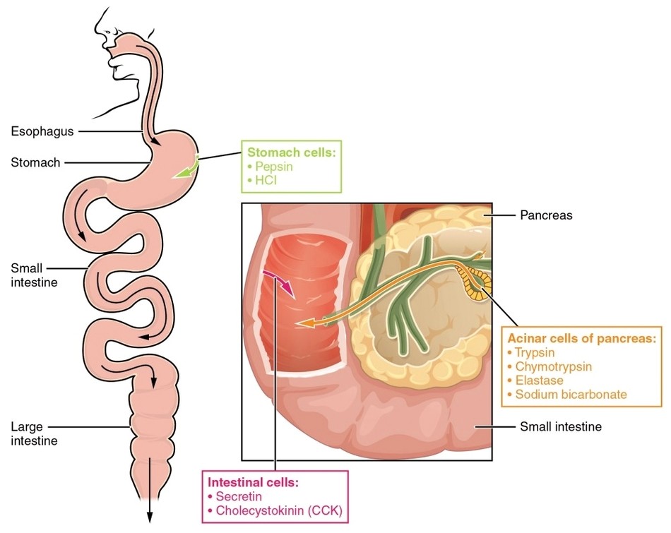
Three Phases Gastric Secretion
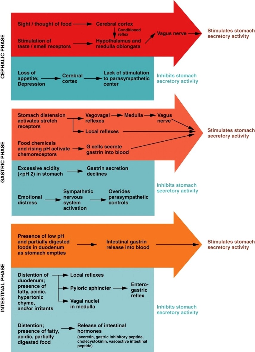
Three Phases Gastric Secretion: Gastric secretion occurs in three phases: the cephalic phase triggered by food anticipation, the gastric phase initiated by stomach distension, and the intestinal phase regulated by chyme entering the intestine.