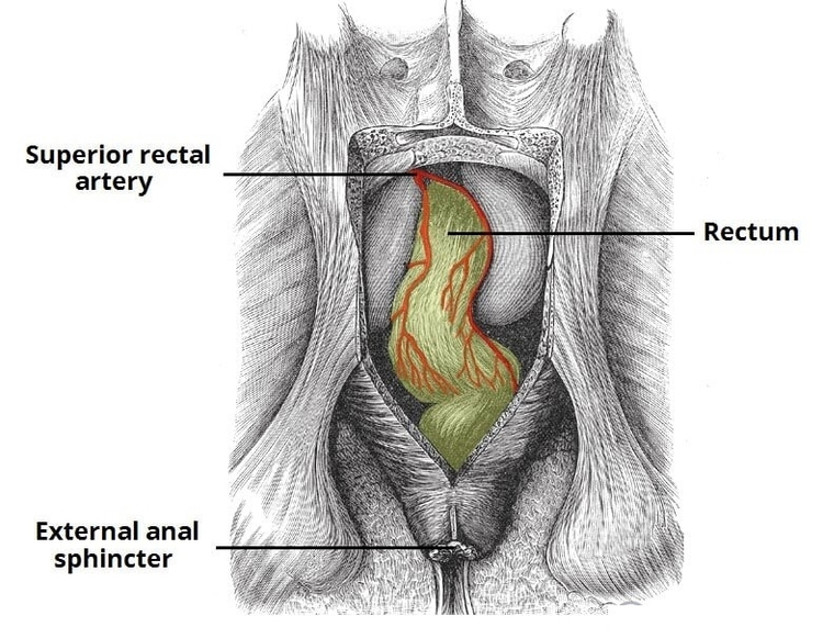
Anatomy Leg By Bk1 Image

72,510 leg anatomy stock photos, vectors, and illustrations are available royalty-free.

When your non-health science friends talk about the ‘leg’, they mean the entire lower extremity. However, in the world of anatomy, the ‘leg’ strictly means the portion between the knee and the ankle joints. It is composed of several structures: In this page, we will take a look at all of the above as well as the anatomy of the knee.

The leg is divided into three major anatomical compartments, each one containing a specific group of muscles: 1 anterior group 2 lateral group 3 posterior group

Anatomy Leg By Bk1 Image Diagram - Chart - diagrams and charts with labels. This diagram depicts Anatomy Leg By Bk1 Image

Anatomy Leg By Bk1 Image

Coloncancer Process Image

The stage refers to the extent of the cancer or how far it has spread. Staging colon cancer is essential to determine the best treatment approach. Colon cancer is typically staged based on a system established by the American Joint Committee on Cancer called the TNM staging system. The system considers the following factors: Primary tumor (T).

Colon cancer 1 Overview. Colon cancer is cancer of the large intestine (colon),… 2 Symptoms. Many people with colon cancer experience no symptoms in the early stages of the disease. 3 Causes. In most cases, it’s not clear what causes colon cancer. 4 Risk factors. The great majority of people diagnosed with colon cancer are older than 50.

After someone is diagnosed with colorectal doctors will try to figure out if it has spread, and if so, how far. This process is called staging. The stage of a cancer describes how much cancer is in the body. It helps determine how serious the cancer is and how best to treat it.

Coloncancer Process Image Diagram - Chart - diagrams and charts with labels. This diagram depicts Coloncancer Process Image

Coloncancer Process Image

Diagram Of Principal Veins Arteries Image

principal veins and arteries. Arteries distribute oxygenated blood throughout the body, while veins carry deoxygenated blood to the heart. Branch of the common iliac artery flowing to the pelvis, the genital organs and the inner thigh. Vein carrying deoxygenated blood from the upper body (above the diaphragm) back to the right atrium.

Here are the steps that the students can follow to draw the arteries and veins. Step 1: at first, people need to draw the heart with the pulmonary artery and vein. The students must place the valves in their proper position, and they can also indicate the four parts of the heart.

Major veins 1 Head and neck. Deoxygenated blood from the brain, head, and neck ultimately drain into one of the three jugular veins: external, internal, or anterior. 2 Trunk. Veins of the trunk converge from the thorax, abdomen, and pelvis towards the heart. … 3 Upper extremity. … 4 Lower extremity. …

Diagram Of Principal Veins Arteries Image Diagram - Chart - diagrams and charts with labels. This diagram depicts Diagram Of Principal Veins Arteries Image

Diagram Of Principal Veins Arteries Image

Diagram Of Week Embryo Image

Your embryo has completed the most critical portion of development. Their skin is still translucent, but their tiny limbs can bend and fine details like nails are starting to form. Read about your pregnancy at 10 weeks.

Your baby moves through different stages, starting as a blastocyst, then maturing into an embryo, and then a fetus. Around the five week mark, your baby’s heart will begin to beat, at 27 weeks they’ll have regular sleep and wake cycles, and at 39 weeks your baby is physically developed.

This page is a link to images from a 10 week female fetus approximately 40 mm in size. This stage of development is after the embryonic period (up to week 8), but only 2 weeks into early fetal development. To see other images from this early fetal stage see Category:Week 10 .

Diagram Of Week Embryo Image Diagram - Chart - diagrams and charts with labels. This diagram depicts Diagram Of Week Embryo Image

Diagram Of Week Embryo Image

Facial Bones Diagram Image

FACIAL BONES ANATOMY. The skull is additionally comprised of fourteen bones which make up the face. The facial bones do not touch the brain but are still considered part of the skull. Some cranial bones meet with the facial bones to give each individual a varying form, the frame work from which the face is then built upon.

Facial bones, or craniofacial bones, are the 14 bones of the skull that articulate with the cranium, protect soft tissues of the face; they aid in breathing, eating, facial expressions, speech, and structure. They are sometimes called the viscerocranium, as they are the bones that make up the face and extend across it.

The facial bones do not touch the brain but are still considered part of the skull. Some cranial bones meet with the facial bones to give each individual a varying form, the frame work from which the face is then built upon.

Facial Bones Diagram Image Diagram - Chart - diagrams and charts with labels. This diagram depicts Facial Bones Diagram Image

Facial Bones Diagram Image

Adam Ear Anatomy Image

Adamimages.com is one of the world’s largest libraries of medical illustrations with nearly 30,000 detailed and medically accurate images ready for immediate download. our comprehensive image catalog covering disease states, injuries, conditions, tests, treatments, surgery procedures and human anatomy.

diagram of the anatomy of the human ear. Three ossicles: malleus, incus, and stapes (hammer, anvil, and stirrup). The ossicles directly couple sound energy from the ear drum to the oval window of the cochlea. Detailed illustration for educational, medical, biological, and scientific use.

The most comprehensive digital database of detailed anatomical images in the world, with the ability to identify over 20,000 anatomical structures from different body orientations (anterior, lateral, medial, posterior, lateral arm, medial arm).

Adam Ear Anatomy Image Diagram - Chart - diagrams and charts with labels. This diagram depicts Adam Ear Anatomy Image

Adam Ear Anatomy Image

Heart Box Diagram Image

The boxes are numbered to correlate with the labeled chambers on the cartoon diagram. Image: Diagram of the heart showing the 4 chambers. We will be using the 4 boxes to represent the chambers of the heart.

We now have a 2×2 table in which we can label the boxes/chambers of the heart. Box 1: The first box is located in the right upper region. We know the atria are on top, and since box 1 is located on the right side, this is the right atrium. Box 2: The second box is also located on the right side, but now we are in the lower region.

Box 1: The first box is located in the right upper region. We know the atria are on top, and since box 1 is located on the right side, this is the right atrium. Box 2: The second box is also located on the right side, but now we are in the lower region.

Heart Box Diagram Image Diagram - Chart - diagrams and charts with labels. This diagram depicts Heart Box Diagram Image

Heart Box Diagram Image

Thyroid Gland Scan Image

An ultrasound is a painless procedure that uses sound waves to generate images of the inside of your body. Your doctor will often use an ultrasound to create images of a fetus during pregnancy. A thyroid ultrasound is used to examine the thyroid for abnormalities, including:

A doctor who specializes in nuclear imaging will evaluate the images and results of your thyroid scan. Your results will be sent in a report to your doctor. A normal thyroid scan would show no abnormalities in the size, shape, and location of the thyroid gland. Your thyroid will have an even green color on the image.

Ultrasound normal thyroid gland has an homogenous appearance, the capsule may appear as a thin hyperechoic line each lobe normally measures 4 length: 4-7 cm volume (excluding isthmus, unless its thickness is >3 mm) 6 10-15 mL for females

Thyroid Gland Scan Image Diagram - Chart - diagrams and charts with labels. This diagram depicts Thyroid Gland Scan Image

Thyroid Gland Scan Image

Asthma Treatment Types Image

Adjust treatment according to your asthma action plan. There are two main types of medications used to treat asthma: Long-term control medications such as inhaled corticosteroids are the most important medications used to keep asthma under control. These preventive medications treat the airway inflammation that leads to asthma symptoms.

Some asthma medicines work quickly to relax your airways and help you breathe easier, while other treatments reduce the swelling and inflammation in your airways and prevent symptoms. It’s important to follow your healthcare provider’s advice about your treatment.

Adjust treatment according to your asthma action plan. Used on a daily basis, these medications can reduce or eliminate asthma flare-ups. Quick-relief inhalers contain a fast-acting medication such as albuterol (Proventil HFA, Ventolin HFA). These medications are sometimes called rescue inhalers.

Asthma Treatment Types Image Diagram - Chart - diagrams and charts with labels. This diagram depicts Asthma Treatment Types Image

Asthma Treatment Types Image

Anatomy Of The Muscles Image

128,412 muscle anatomy stock photos, vectors, and illustrations are available royalty-free.

Forearms – Anatomy Muscles Rhomboid minor and rhomboid major, levator scapulae and latissimus dorsi muscles – didactic board of anatomy of human bony and muscular system, posterior view Abs – Anatomy Muscles Leg muscles of the man Human Body Organs (Lungs) 3D Triceps – Anatomy Muscles Hand muscle connection with brain

Last Updated: Jul 16, 2019 The muscular system is responsible for the movement of the human body. Attached to the bones of the skeletal system are about 700 named muscles that make up roughly half of a person’s body weight. Each of these muscles is a discrete organ constructed of skeletal muscle tissue, blood vessels, tendons, and nerves.

Anatomy Of The Muscles Image Diagram - Chart - diagrams and charts with labels. This diagram depicts Anatomy Of The Muscles Image

Anatomy Of The Muscles Image

Dep The Abo Blood Group Diagram Image

It is not to be confused with Typo or Type 0. The ABO blood group system is used to denote the presence of one, both, or neither of the A and B antigens on erythrocytes. In human blood transfusions it is the most important of the 38 different blood type (or group) classification systems currently recognized.

ABO antibodies in the serum are formed naturally. when the immune system encounters the “missing” ABO blood group antigens in foods or in micro-organisms. This happens at an early age because sugars that are identical to, or very similar to, the ABO blood group antigens are found

The four basic ABO phenotypes are O, A, B, and AB. After it was found that blood group A RBCs reacted differently to a particular antibody (later called anti-A1), the blood group was divided into two phenotypes, A 1 and A 2.

Dep The Abo Blood Group Diagram Image Diagram - Chart - diagrams and charts with labels. This diagram depicts Dep The Abo Blood Group Diagram Image

Dep The Abo Blood Group Diagram Image

Gdm Risk Factors Image

The US Diabetes Prevention Program included women with a history of GDM as one risk factor for diabetes mellitus.

With obesity becoming a global epidemic, the assessment of the strength of the evidence supporting the impact of overweight and obesity in GDM could allow the identification of women at high risk for adverse outcomes and allow better prevention.

A hyperglycemic environment is associated with oxidative stress, and GDM women have been reported to overproduce free radicals and have impaired free-radical scavenging mechanisms [139]. ROS inhibit insulin-stimulated glucose uptake by interfering with both IRS-1 and GLUT4 [140].

Gdm Risk Factors Image Diagram - Chart - diagrams and charts with labels. This diagram depicts Gdm Risk Factors Image

Gdm Risk Factors Image

Bones In Human Body Image

67,060 human bone stock photos and images available, or search for human bone marrow or human bone structure to find more great stock photos and pictures.

Human skeleton hand, knee and shoulder, hip, foot, spine, leg and arm, finger, elbow, pelvis, thorax, ankle, wrist icon for orthopedics and rheumatology medicine design Layer of a Long bone. Humerus. Tube. Human bones skeleton silhouette collection set.

The skeleton makes up about 30-40% of an adult’s body mass. The skeleton’s mass is made up of nonliving bone matrix and many tiny bone cells. Roughly half of the bone matrix’s mass is water, while the other half is collagen protein and solid crystals of calcium carbonate and calcium phosphate.

Bones In Human Body Image Diagram - Chart - diagrams and charts with labels. This diagram depicts Bones In Human Body Image

Bones In Human Body Image

Anatomy Arm By Bk Image

58,682 arm anatomy stock photos, vectors, and illustrations are available royalty-free. See arm anatomy stock video clips

The human arm is divided into two main regions, the portion from the elbow to the wrist known as the forearm, and the segment from the shoulder to the elbow referred to as the arm. Arm muscle anatomy enables the arm to perform a variety of movements, including flexion, extension, pronation, and supination.

The (upper) arm muscles are a group of five muscles located in the region between the shoulder and elbow joints. They are divided into two distinct compartments of the arm.

Anatomy Arm By Bk Image Diagram - Chart - diagrams and charts with labels. This diagram depicts Anatomy Arm By Bk Image

Anatomy Arm By Bk Image

Broken Foot Ray Sized Image

However, even with an X-ray it can be difficult to determine if your foot is broken as swelling can obscure the fine bones in the foot. Using an X-ray, the doctor may be able to identify which bones in your foot are broken and how they can be treated.

The major symptom of a broken foot is overwhelming pain when you try to put any pressure at all on your foot or walk on your foot. If you have a broken toe, you can usually still walk and not be in too much pain.

Human foot ankle and leg in x-ray, on gray background. The foot ankle is highlighted by green colour X-ray ankle show fracture distal tibia and fibula (leg’s bone). Film x-ray ankle show fracture distal tibia and fibula (leg’s bone

Broken Foot Ray Sized Image Diagram - Chart - diagrams and charts with labels. This diagram depicts Broken Foot Ray Sized Image

Broken Foot Ray Sized Image

Calf Muscle Diagram Image

The calf is defined largely as the three headed calf muscle (triceps surae muscle). In anatomy, we actually talk about the lower leg muscles and divide them into the following categories:

Picture of the Calf Muscle. The gastrocnemius is the larger calf muscle, forming the bulge visible beneath the skin. The gastrocnemius has two parts or “heads,” which together create its diamond shape. The soleus is a smaller, flat muscle that lies underneath the gastrocnemius muscle.

Calf muscle ultrasound: A device placed on the skin bounces high-frequency sound waves off the calf muscle, tendon, and other structures in the leg. The signals are converted into images on a video screen, allowing health-care providers to see structures inside the body. 1 2

Calf Muscle Diagram Image Diagram - Chart - diagrams and charts with labels. This diagram depicts Calf Muscle Diagram Image

Calf Muscle Diagram Image