Diagram Foot Anatomy Mw Image

This human anatomy diagram with labels depicts and explains the details and or parts of the Diagram Foot Anatomy Mw Image. Human anatomy diagrams and charts show internal organs, body systems, cells, conditions, sickness and symptoms information and/or tips to ensure one lives in good health.

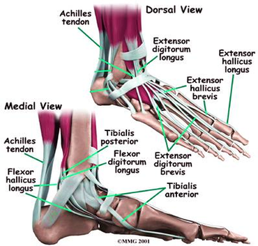
The foot diagram has a complex structure made up of bones, ligaments, muscles, and tendons. Understanding the structure of the foot is best done by looking at a foot diagram where the anatomy has been labeled.

The anatomy of the foot. The foot contains a lot of moving parts – 26 bones, 33 joints and over 100 ligaments. The foot is divided into three sections – the forefoot, the midfoot and the hindfoot. The forefoot. This consists of five long metatarsal bones and five shorter bones that form the toes (phalanges).

The medial column is more mobile and consists of the talus, navicular, medial cuneiform, 1st metatarsal, and great toe. The lateral column is stiffer and includes the calcaneus, cuboid, and the 4th and 5th metatarsals. Figure 3: Columns of the Foot

Diagram Foot Anatomy Mw Image

Diagram Foot Anatomy Mw Image


Tags: ,