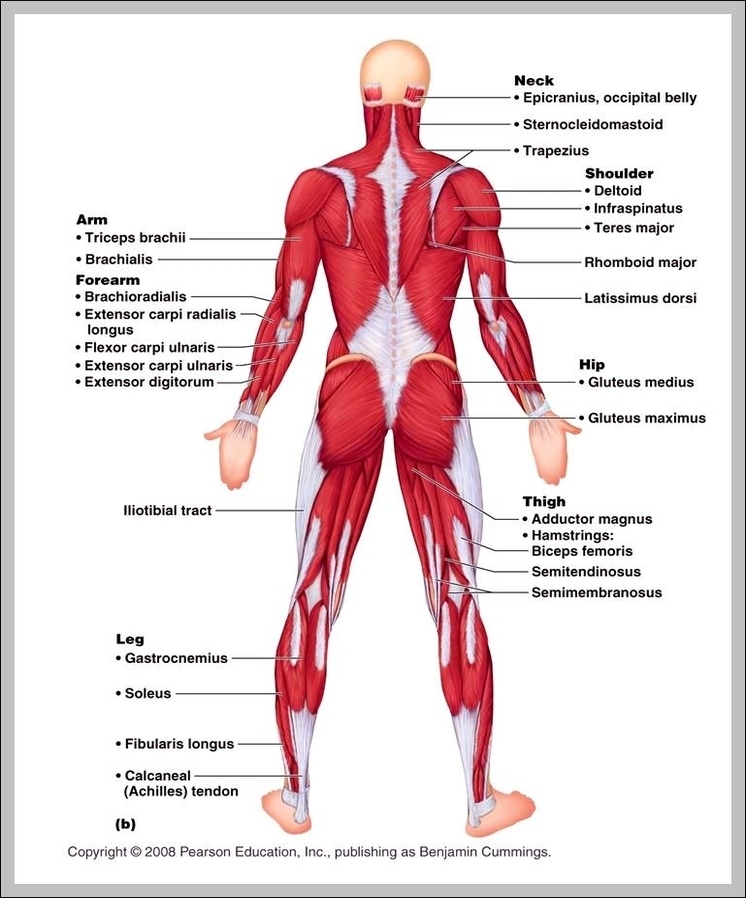
Upper Back Muscle Anatomy
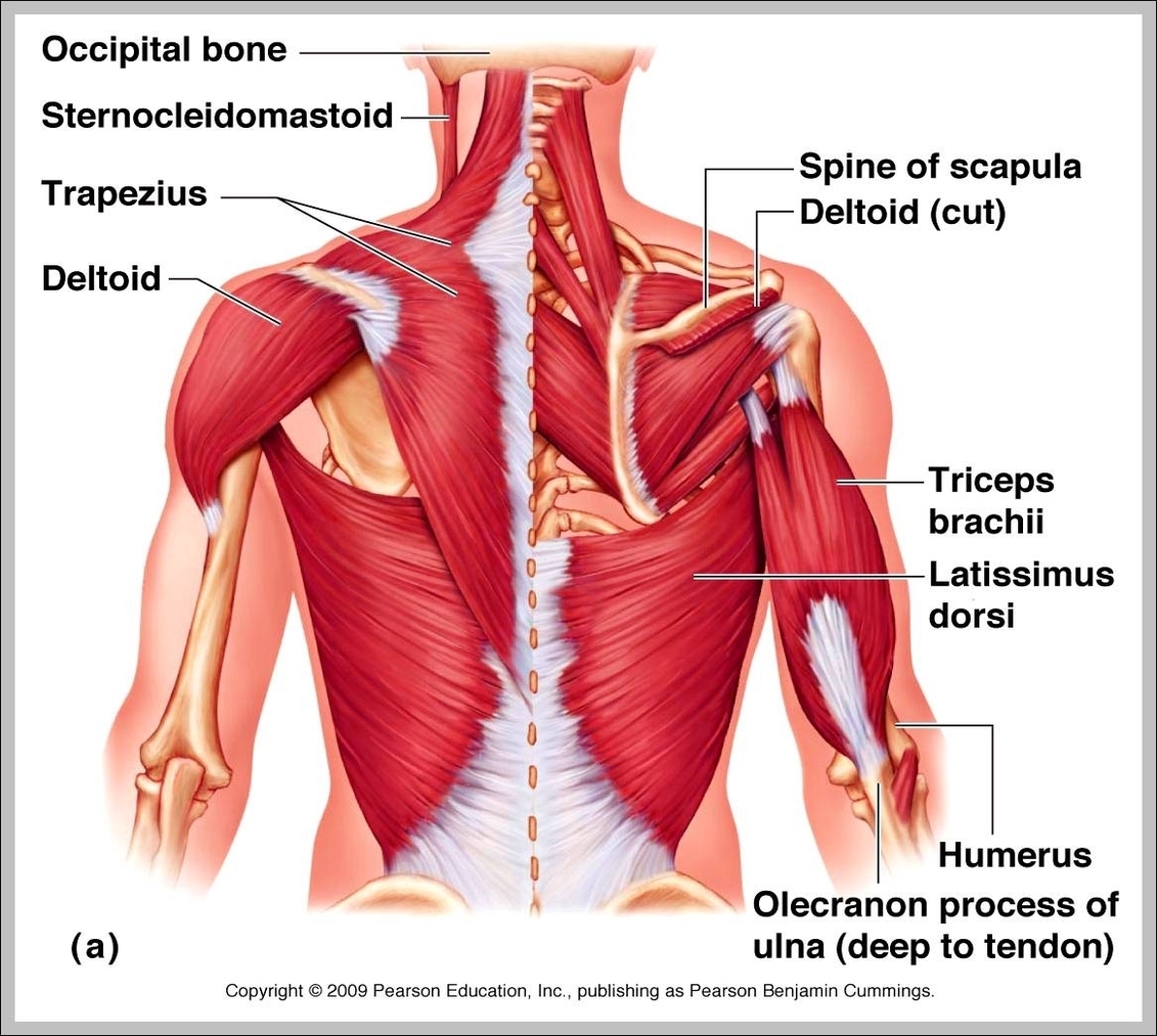
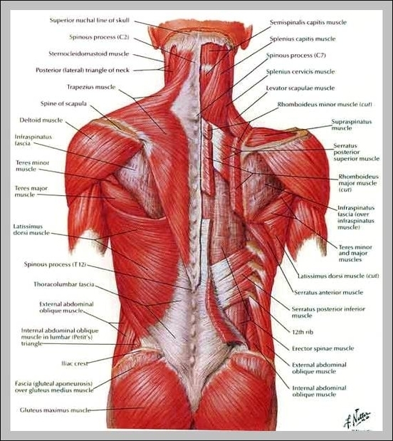
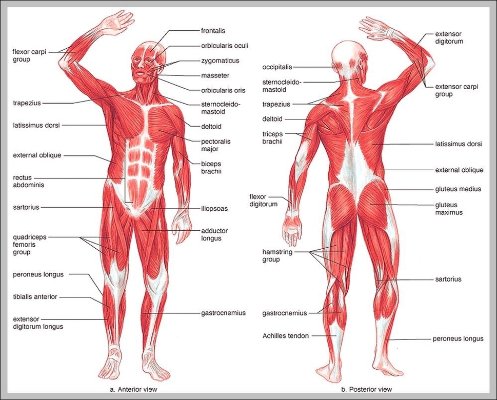
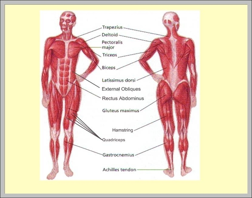
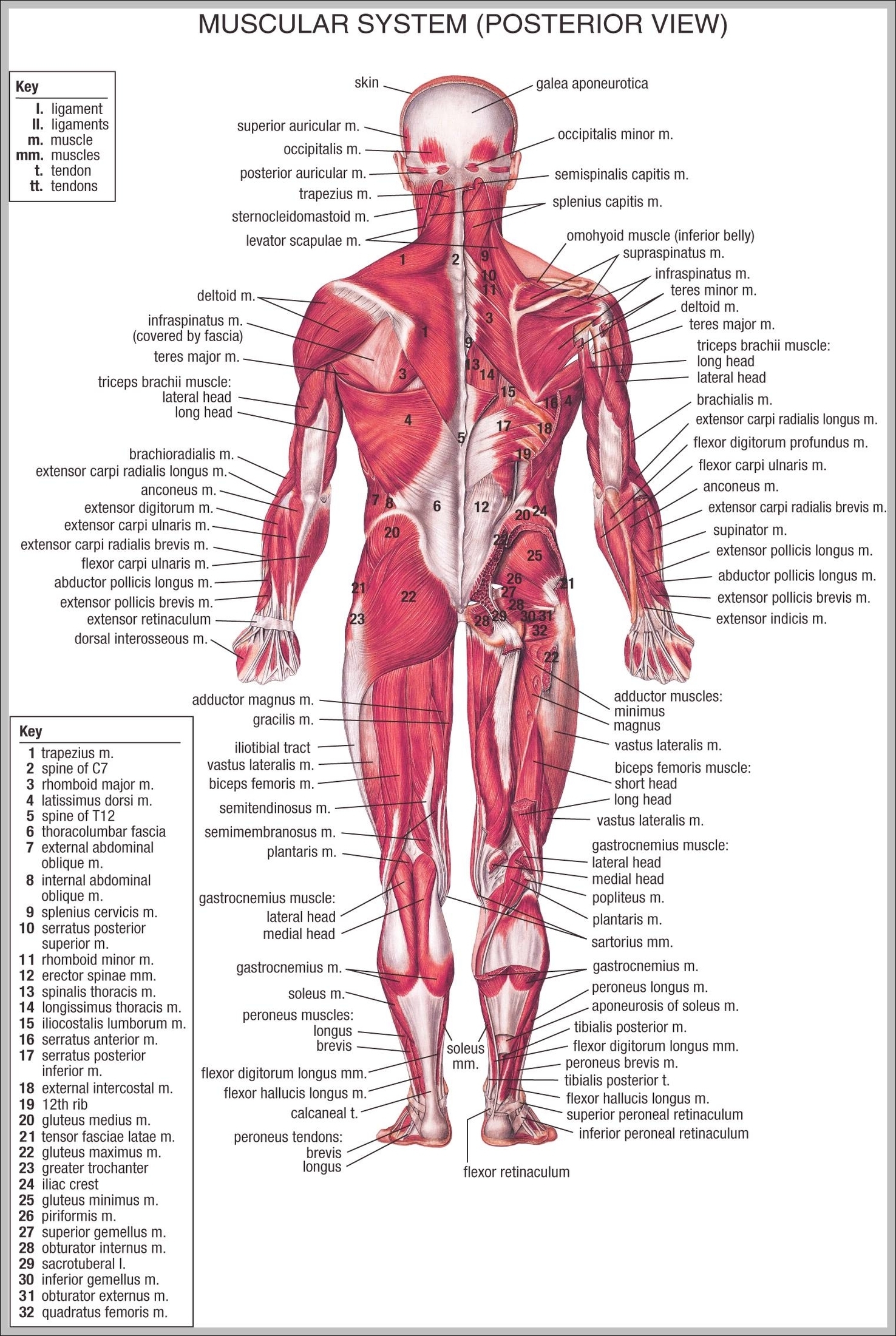
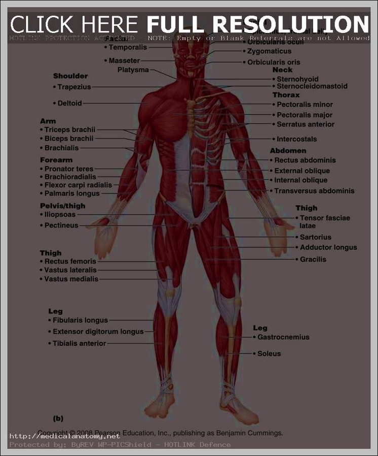
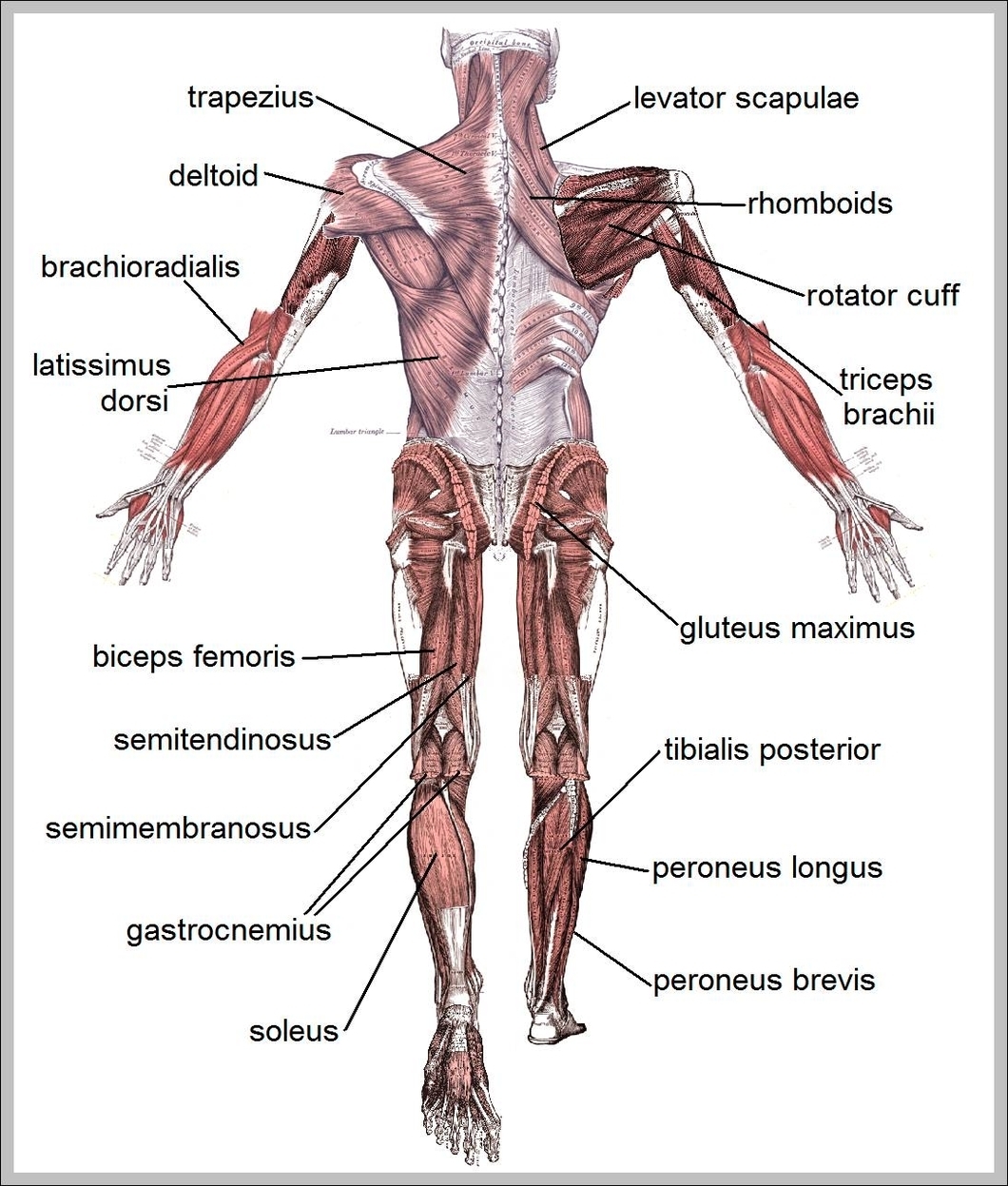
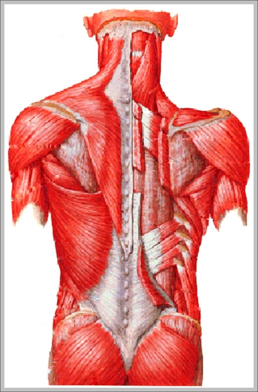
Upper Back Muscle Anatomy: The upper back muscles include the trapezius, rhomboids, and latissimus dorsi, all of which help with posture, shoulder movement, and arm extension.