
Bones Of The Ankle
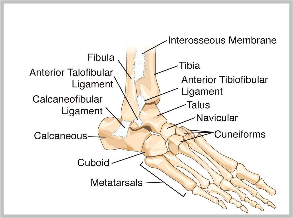
Bones Of The Ankle: The ankle is composed of the tibia, fibula, and talus, along with smaller bones like the calcaneus and navicular, which form a flexible and stable joint.
Bones Of The Ankle: The ankle is composed of the tibia, fibula, and talus, along with smaller bones like the calcaneus and navicular, which form a flexible and stable joint.
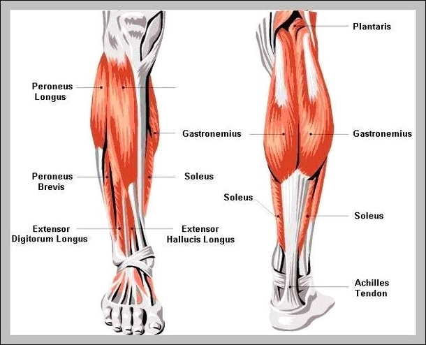
Bones Of Lower Leg: The lower leg contains the tibia (shinbone) and fibula, which support weight, enable movement, and connect to the knee and ankle joints.
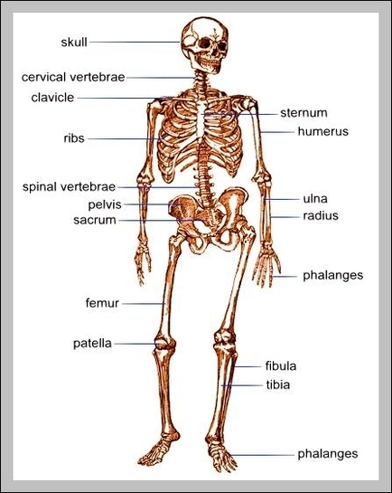
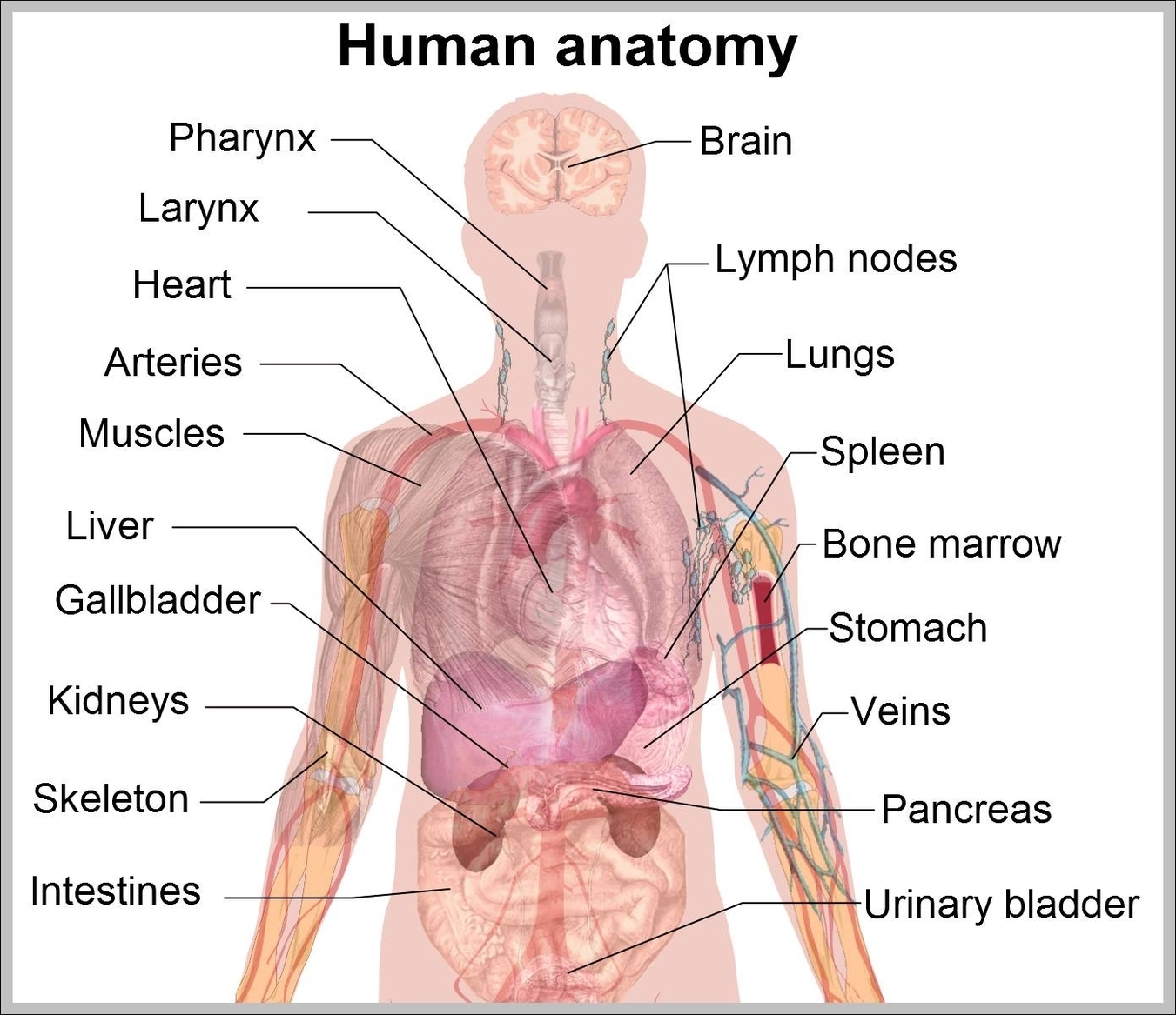
Bones Of Human Body: The human body has 206 bones that form the skeletal system. These bones provide structure, protect vital organs, store minerals, and enable movement. Major bones include the skull, spine, ribs, arms, and legs.
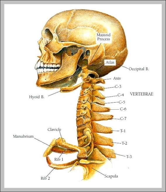
Bones In Your Neck: The neck contains seven cervical vertebrae, supporting the skull and enabling neck movement. Ligaments and muscles surround these bones, helping with stability and movement.
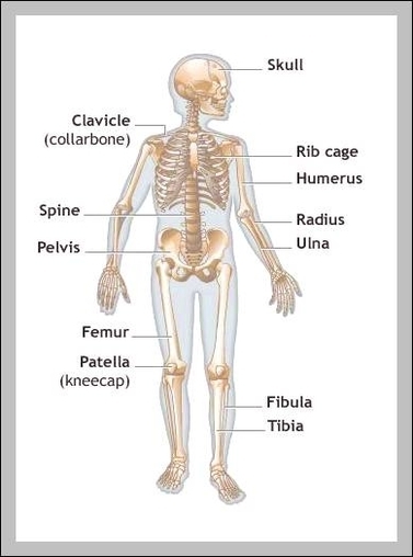
Bones In Your Legs: The bones in the legs include the femur, tibia, fibula, and patella. These bones provide structure, allow movement, and bear the weight of the body during activities like walking and running.
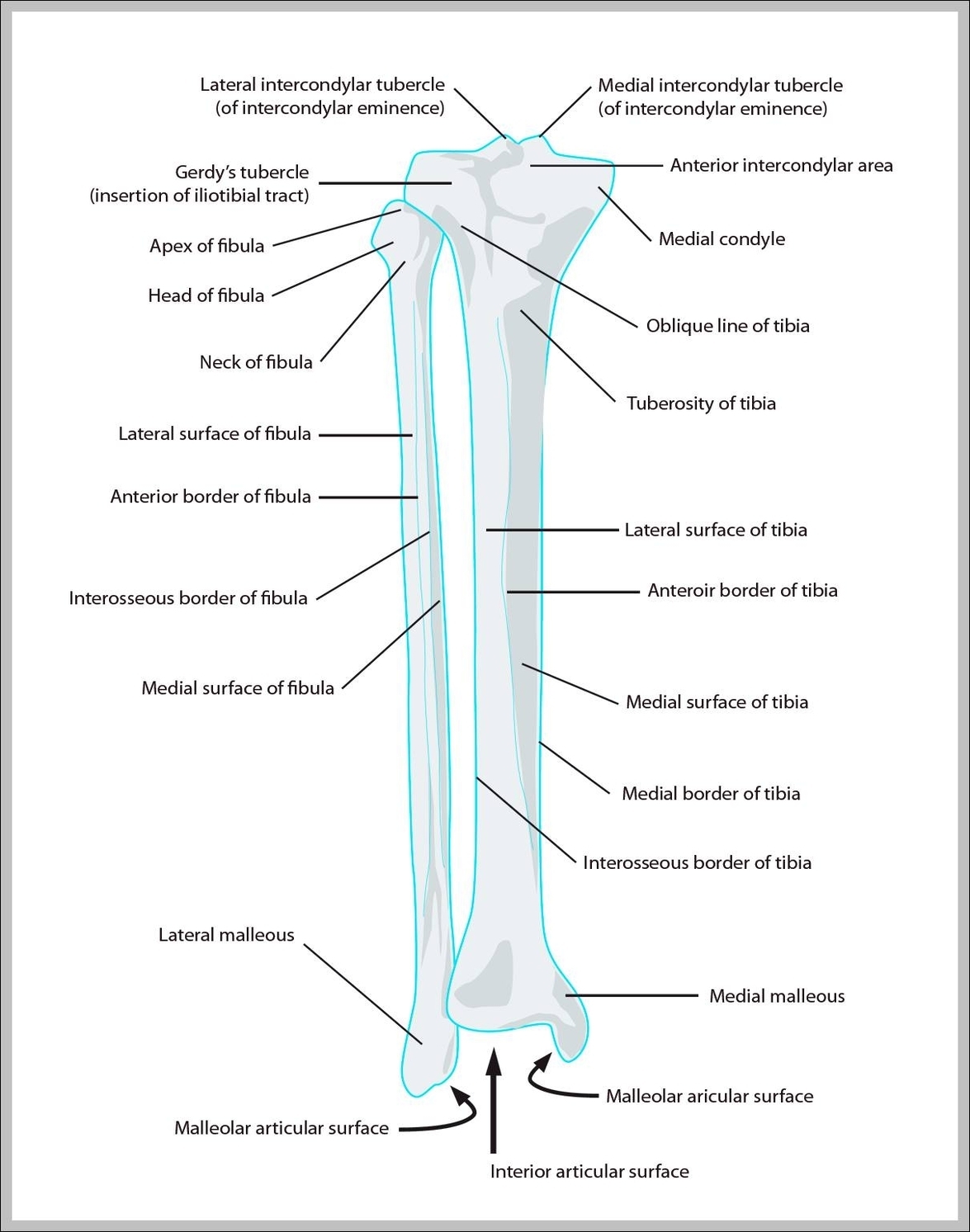
Bones In The Lower Leg: The bones in the lower leg include the tibia and fibula, which support body weight and facilitate movement.
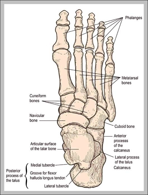
Bones In Feet: The bones in the feet include the tarsals, metatarsals, and phalanges. They provide the structure and support needed for standing, walking, and running, distributing weight and absorbing shock.
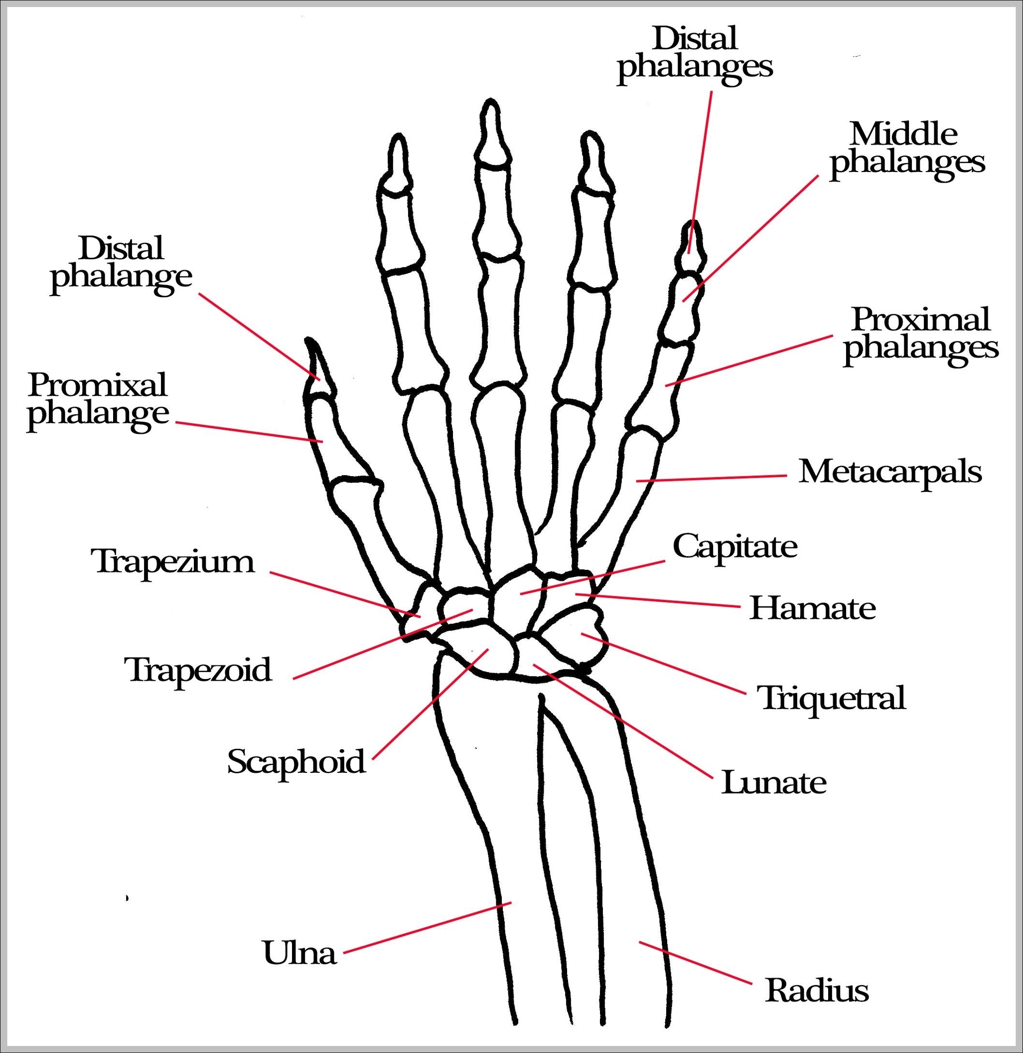
Bones Hand: The bones of the hand include the phalanges, metacarpals, and carpals, which together allow for complex movements and dexterity. The hand’s bones support grasping, fine motor skills, and gestures.
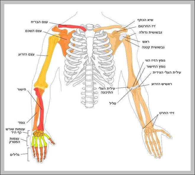
Arms Bones: The arm bones include the humerus (upper arm), radius and ulna (forearm), and associated joints, enabling flexibility and strength for lifting and fine motor tasks.

Arm And Hand Bones: The bones in the arm and hand include the humerus, radius, ulna, carpals, metacarpals, and phalanges. These bones support movement, strength, and dexterity in upper limbs.
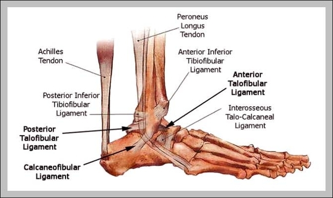
Ankle Bones Diagram: An ankle bones diagram shows the tibia, fibula, and tarsal bones, which make up the structure of the ankle and allow for movement.
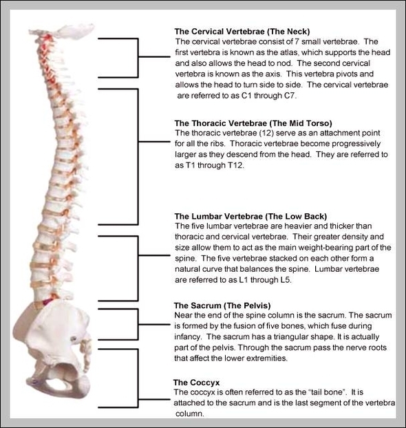
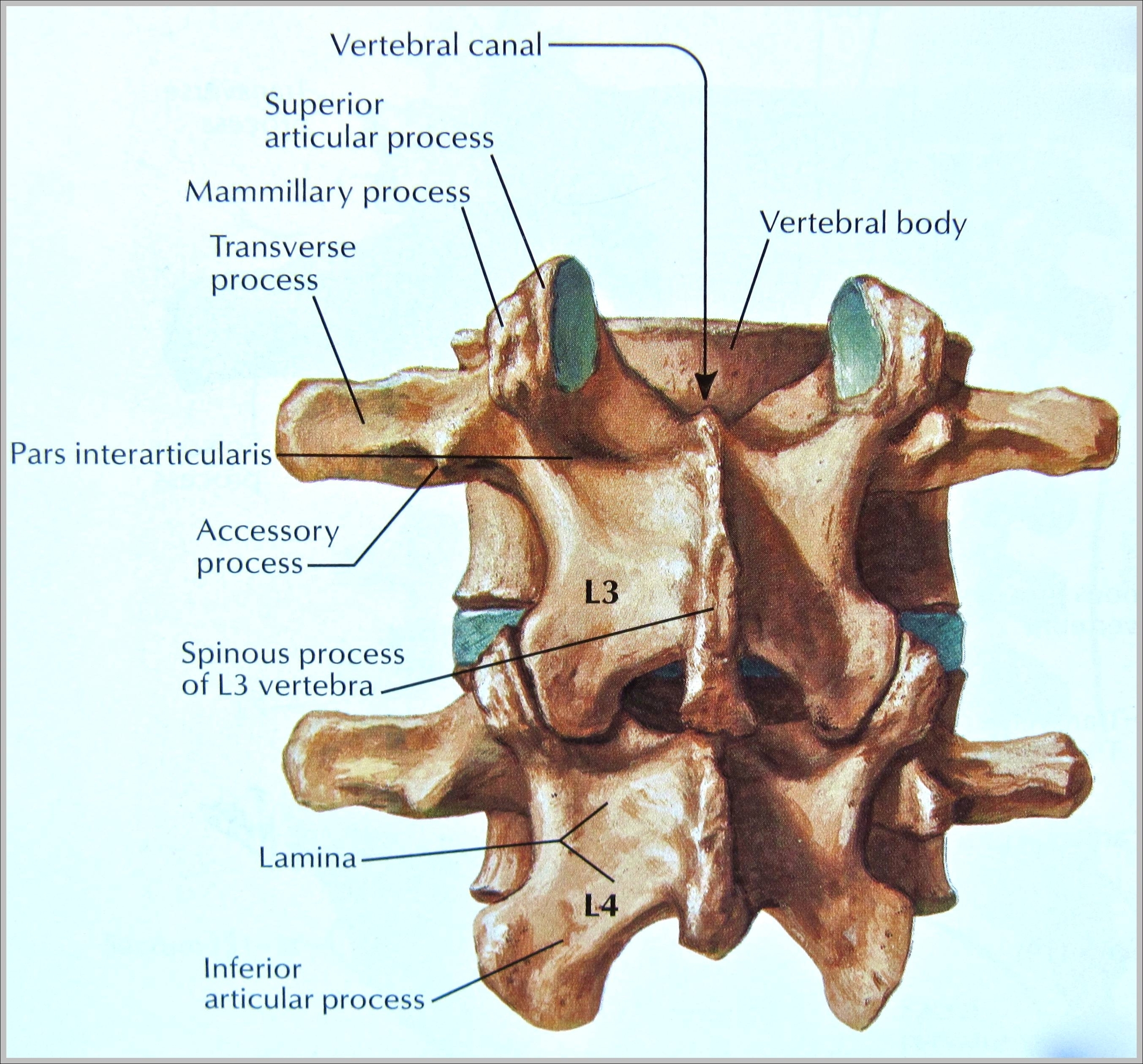
Anatomy Of Vertebrae: Vertebral anatomy includes the vertebral body, arch, spinous process, and spinal canal. These structures protect the spinal cord and support posture and movement.
Anatomy Of Skull Bones: The skull consists of 22 bones divided into cranial bones (protecting the brain) and facial bones (forming the face), including the frontal, parietal, occipital, temporal, and mandible.
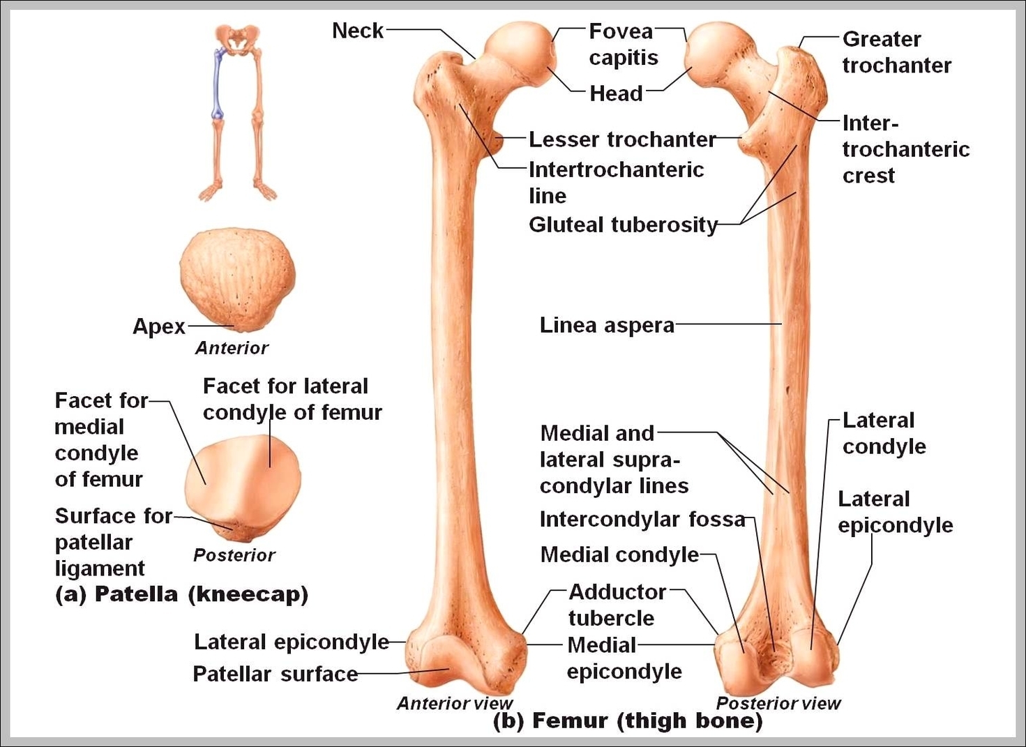
Anatomy Of Femur: The femur is the thigh bone and the longest and strongest bone in the human body. It connects the hip to the knee and is crucial for weight-bearing and movement.
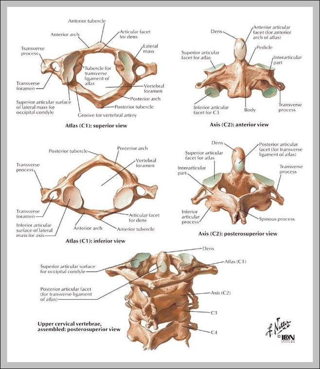
Anatomy Of Cervical Vertebrae: The cervical vertebrae are the seven spinal bones in the neck region. They support the head, protect the spinal cord, and allow for a wide range of neck motion.
3rd Vertebrae: The third cervical vertebra (C3) supports neck mobility and spinal cord protection, lying between C2 and C4. It plays a critical role in head and neck function.

2nd Vertebrae: The second vertebra, or axis (C2), supports the atlas (C1) and enables the head to rotate from side to side, forming a key part of the cervical spine.