Diagram Of Dental Cavity Image
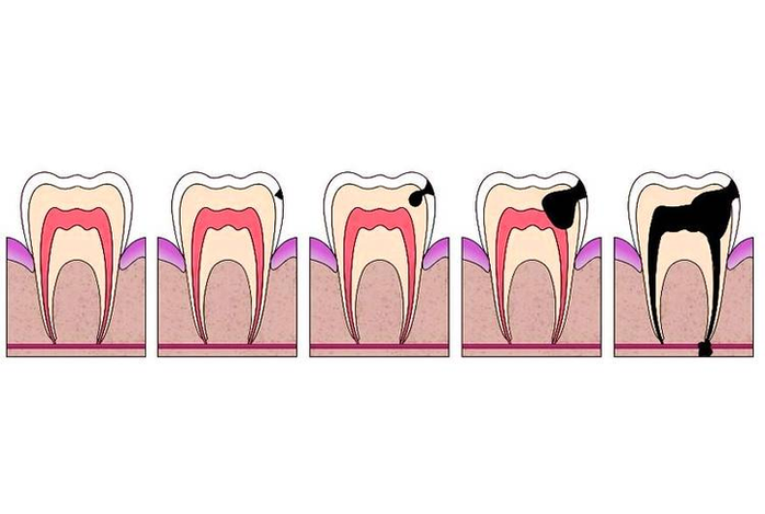
69,050 oral cavity stock photos, vectors, and illustrations are available royalty-free. 38 nasal cavity diagram stock photos and images available or start a new search to explore more stock photos and images. What is the Oral Cavity. The human oral View Diagram Diagram Of Dental Cavity Image