Testis Diagram Image

This human anatomy diagram with labels depicts and explains the details and or parts of the Testis Diagram Image. Human anatomy diagrams and charts show internal organs, body systems, cells, conditions, sickness and symptoms information and/or tips to ensure one lives in good health.

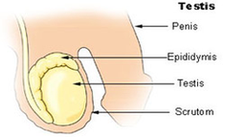
58 testes diagram stock photos and images available, or start a new search to explore more stock photos and images. The Principal Glands Of The Female And Male Human Endocrine Systems. An anatomical diagram showing the muscular and cutaneous distribution of the small sciatic or inferior gluteal nerve, 1893.

What are testes? The testes — also called testicles — are two oval-shaped organs in the male reproductive system. They’re contained in a sac of skin called the scrotum. The scrotum hangs outside the body in the front of the pelvic region near the upper thighs.

The testes are connected to the vital organs of the ventral body cavity via the spermatic cords. Nerves, blood vessels, and lymphatic vessels travel through the spermatic cords to support the testes. The vas deferens also passes through the spermatic cord carrying sperm out of the testes toward the prostate and urethra.

Testis Diagram Image

Testis Diagram Image


Tags: