
Hiv Transmission Electron Micrograph Aidsbbb Lores Figure Image
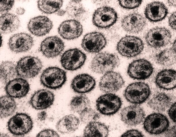
Through the use of advanced scanning electron microscopy (SEM) and other imaging techniques, scientists have a far greater ability to investigate the ultrastructure of HIV and other infective microbes related to HIV diseases. A scanning electron micrograph of a human View Diagram Hiv Transmission Electron Micrograph Aidsbbb Lores Figure Image