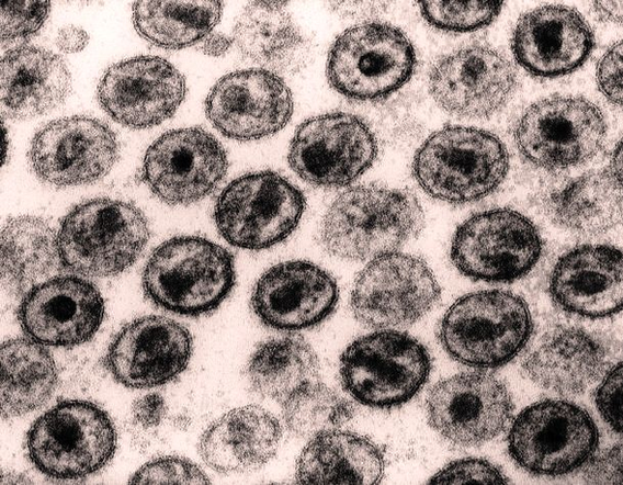
Through the use of advanced scanning electron microscopy (SEM) and other imaging techniques, scientists have a far greater ability to investigate the ultrastructure of HIV and other infective microbes related to HIV diseases. A scanning electron micrograph of a human T-lymphocyte (also called a T-cell) from the immune system of a healthy donor. Hiv Transmission Electron Micrograph Aidsbbb Lores Figure Image Diagram - Chart - diagrams and charts with labels. This diagram depicts Hiv Transmission Electron Micrograph Aidsbbb Lores Figure Image and explains the details of Hiv Transmission Electron Micrograph Aidsbbb Lores Figure Image.
Hiv Transmission Electron Micrograph Aidsbbb Lores Figure Image