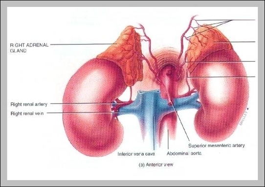
Picture Of Adrenal Glands

This human anatomy diagram with labels depicts and explains the details and or parts of the Picture Of Adrenal Glands. Human anatomy diagrams and charts show internal organs, body systems, cells, conditions, sickness and symptoms information and/or tips to ensure one lives in good health.

Anatomical Structure The adrenal glands consist of an outer connective tissue capsule, a cortex and a medulla. Veins and lymphatics leave each gland via the hilum, but arteries and nerves enter the glands at numerous sites. The outer cortex and inner medulla are the functional portions of the gland.

The suprarenal gland (adrenal gland) is located at the top of each kidney; therefore each person has two suprarenal glands. The suprarenal glands are divided into two parts. The outer portion of the gland is called the adrenal cortex.

Some of the popular cancer cells like lung cancer, melanoma, and breast cancer can finds its path to reach adrenal gland. Cancer or adrenal glands tumor is formed from adrenal cortex region or from adrenal medulla. In rare cases, lymphoma cancer can occur on the adrenal gland.

Picture Of Adrenal Glands

picture of adrenal glands