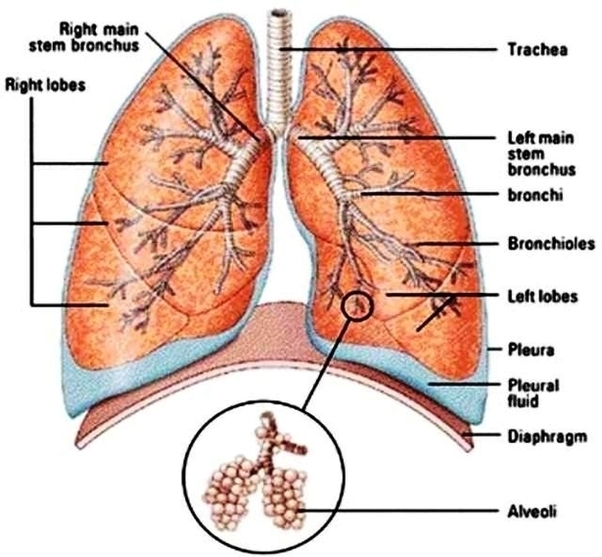
Lung Diagram Small Image

This human anatomy diagram with labels depicts and explains the details and or parts of the Lung Diagram Small Image. Human anatomy diagrams and charts show internal organs, body systems, cells, conditions, sickness and symptoms information and/or tips to ensure one lives in good health.

8,253 lung diagram stock photos, vectors, and illustrations are available royalty-free.

A healthy lung has a pinkish appearance, and if you could see it outside the body, it would look like a squishy, rubbery structure [4]. The lungs are located a little toward the posterior part of the human body, just below the collarbone, extending down to the diaphragm, the muscular partition that separates the chest and abdominal cavities.

What are Lungs: Definition. The spongy air-filled conical organs occupying most of the thoracic (chest) cavity in humans are known as the lungs [1]. It is one of the primary respiratory organs where the gas exchange occurs after the inhaled air enters the lungs via the trachea, through the bronchi and bronchioles [2].

Lung Diagram Small Image

Lung Diagram Small Image


Tags: ,