Knee joint is one of the most important hinge joints of our body. Its complexity and its efficiency is the best example of God’s creation. The anatomy of the knee consists of bones, muscles, nerves, cartilages, tendons and ligaments. All these parts combine and work together. Knee Diagram Tendons Image Diagram - Chart - diagrams and charts with labels. This diagram depicts Knee Diagram Tendons Image and explains the details of Knee Diagram Tendons Image.
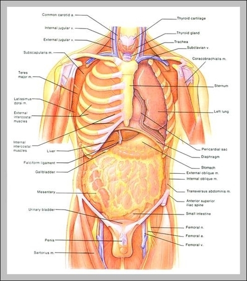
Knee Diagram Tendons Image