Human Skin Anatomy

This human anatomy diagram with labels depicts and explains the details and or parts of the Human Skin Anatomy. Human anatomy diagrams and charts show internal organs, body systems, cells, conditions, sickness and symptoms information and/or tips to ensure one lives in good health.

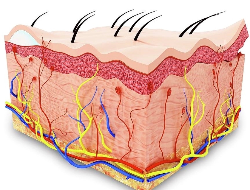
Human skin, in human anatomy, the covering, or integument, of the body’s surface that both provides protection and receives sensory stimuli from the external environment. The skin consists of three layers of tissue: the epidermis, an outermost layer that contains the primary protective structure,

Skin has three layers: The epidermis, the outermost layer of skin, provides a waterproof barrier and creates our skin tone. The dermis, beneath the epidermis, contains tough connective tissue, hair follicles, and sweat glands. The deeper subcutaneous tissue (hypodermis) is made of fat and connective tissue.

It’s located between the epidermis and the subcutaneous tissue. It contains connective tissue, blood capillaries, oil and sweat glands, nerve endings, and hair follicles. The dermis is split into two parts—the papillary dermis, which is the thin, upper layer, and the reticular dermis, which is the thick, lower layer. 5

Human Skin Anatomy

Human skin anatomy


Tags: , ,