Pictures Of The Endocrine System
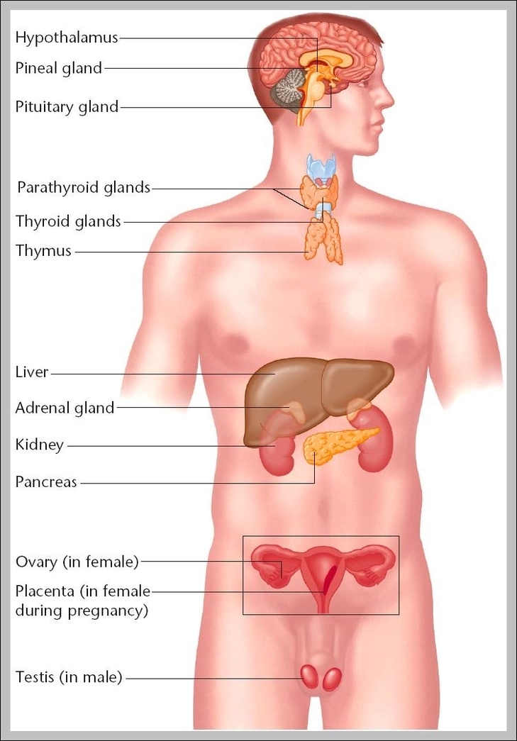
Pictures Of The Endocrine System: Pictures of the endocrine system typically highlight glands like the pituitary, thyroid, adrenal, pancreas, and reproductive glands, showing hormone-producing organs throughout the body.