Autonomic Nervous System Image

This human anatomy diagram with labels depicts and explains the details and or parts of the Autonomic Nervous System Image. Human anatomy diagrams and charts show internal organs, body systems, cells, conditions, sickness and symptoms information and/or tips to ensure one lives in good health.

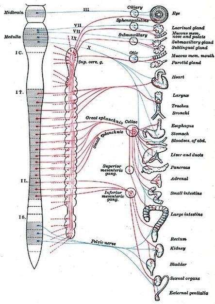
Autonomic nervous system (ANS) is a functional division of the nervous system, with its structural parts in both the central nervous system (CNS) and the peripheral nervous system (PNS). It controls the glands and smooth muscle of all the internal organs (viscera) unconsciously.

The hypothalamus, just above the brain stem, acts as an integrator for autonomic functions, receiving ANS regulatory input from the limbic system to do so. The autonomic nervous system has three branches: the sympathetic nervous system, the parasympathetic nervous system and the enteric nervous system.

The autonomic nervous system is unique in that it requires a sequential two-neuron efferent pathway; the preganglionic neuron must first synapse onto a postganglionic neuron before innervating the target organ.

Autonomic Nervous System Image

Autonomic Nervous System Image


Tags: , ,