Skeletal Muscles Image Diagram - Chart - diagrams and charts with labels. This diagram depicts Skeletal Muscles Image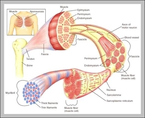
Category Archives: Muscles
Muscles System Image
98 muscular system labeled diagram stock photos and images available, or start a new search to explore more stock photos and images. Frontal view of the muscular system of the male human body with descriptive labels pointing to the muscles on a white background. Labeled Anatomy Chart of Neck and Shoulder Muscles on White…
Last Updated: Jul 16, 2019 The muscular system is responsible for the movement of the human body. Attached to the bones of the skeletal system are about 700 named muscles that make up roughly half of a person’s body weight. Each of these muscles is a discrete organ constructed of skeletal muscle tissue, blood vessels, tendons, and nerves.
Human muscle system, the muscles of the human body that work the skeletal system, that are under voluntary control, and that are concerned with movement, posture, and balance. Broadly considered, human muscle—like the muscles of all vertebrates—is often divided into striated muscle (or skeletal muscle), smooth muscle, and cardiac muscle.
Muscles System Image Diagram - Chart - diagrams and charts with labels. This diagram depicts Muscles System Image
Shoulder Back Muscles Image
These functions include: Trapezius: Your traps serve to elevate your shoulders. The middle and lower trapezius function to retract your shoulders, pulling them backward. Rhomboids: The rhomboids serve to retract and stabilize your shoulder blades. Latissimus dorsi: Your latissimus functions to extend and medially rotate your upper arm bone.
124,600 shoulder muscles stock photos, vectors, and illustrations are available royalty-free. See shoulder muscles stock video clips
Working on the pectoral girdle, the trapezius, rhomboid major, and levator scapulae muscles of the back elevate the scapula to shrug the shoulders and move the scapula posteriorly (as in reaching back behind the body).
Shoulder Back Muscles Image Diagram - Chart - diagrams and charts with labels. This diagram depicts Shoulder Back Muscles Image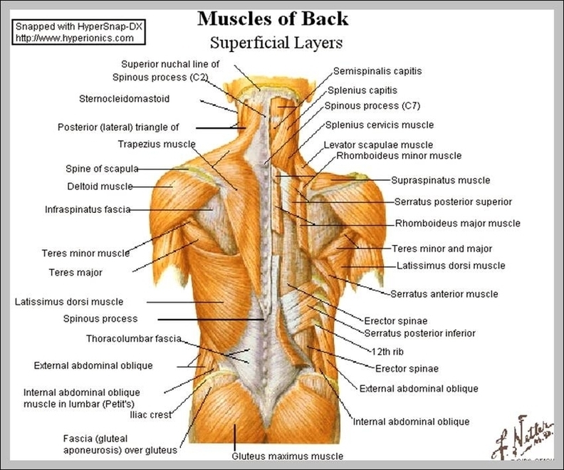
Muscles Of The Back Diagram Image
There are three different muscle groups found in the back: the superficial group, the deep group, and the intermediate group. Muscles found in the superficial group include rhomboid major, rhomboid minor, levator scapulae, trapezius, latissimus dorsi.
25,649 back muscle anatomy stock photos, vectors, and illustrations are available royalty-free.
The back supports the weight of the body, allowing for flexible movement while protecting vital organs and nerve structures. This article looks at the anatomy of the back, including bones, muscles, and nerves. It also covers some common conditions and injuries that can affect the back.
Muscles Of The Back Diagram Image Diagram - Chart - diagrams and charts with labels. This diagram depicts Muscles Of The Back Diagram Image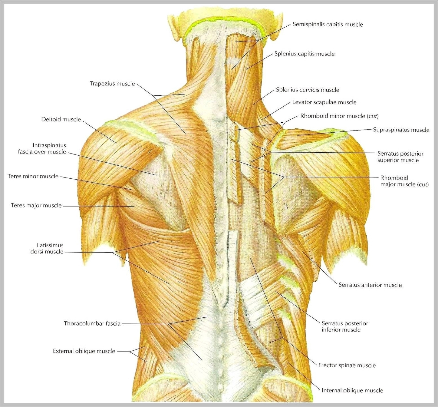
Human Muscle Model Image
105,188 human muscle anatomy stock photos, vectors, and illustrations are available royalty-free.
Forearms – Anatomy Muscles Rhomboid minor and rhomboid major, levator scapulae and latissimus dorsi muscles – didactic board of anatomy of human bony and muscular system, posterior view Abs – Anatomy Muscles Leg muscles of the man Human Body Organs (Lungs) 3D Triceps – Anatomy Muscles Hand muscle connection with brain
Last Updated: Jul 16, 2019 The muscular system is responsible for the movement of the human body. Attached to the bones of the skeletal system are about 700 named muscles that make up roughly half of a person’s body weight. Each of these muscles is a discrete organ constructed of skeletal muscle tissue, blood vessels, tendons, and nerves.
Human Muscle Model Image Diagram - Chart - diagrams and charts with labels. This diagram depicts Human Muscle Model Image
Muscle Bodies Image
Muscle Bodies Image Diagram - Chart - diagrams and charts with labels. This diagram depicts Muscle Bodies Image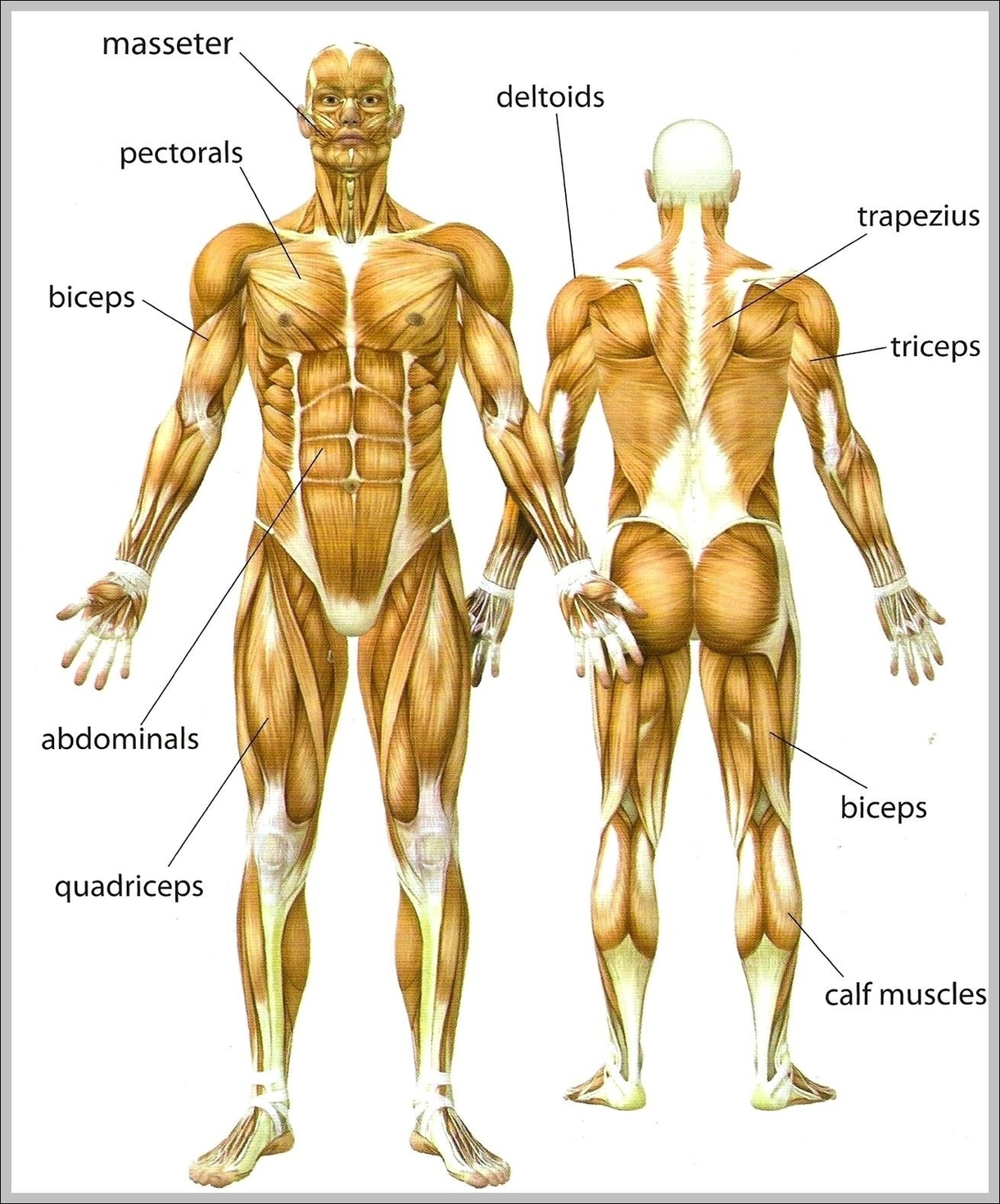
Anatomy And Physiology Of Animals Structure Of Muscle Image
Here is a nice 50-questions quiz on animal muscles both as gross anatomy and in a microscope. This quiz; however, will focus on the gross anatomy of muscles.
Muscle is a soft tissue found in most animals. They are primarly responsible for maintaining and changing posture,locomotion as well as movement of internal organs. They are derived from the mesodermal layer of embryonic germ cells in a process known as myogenesis.
Different skeletal muscles in animals are described as white and red muscle. These different types of skeletal muscles are recruited depending on whether a fast and short versus steady and prolonged locomotion is needed by the animal.
Anatomy And Physiology Of Animals Structure Of Muscle Image Diagram - Chart - diagrams and charts with labels. This diagram depicts Anatomy And Physiology Of Animals Structure Of Muscle Image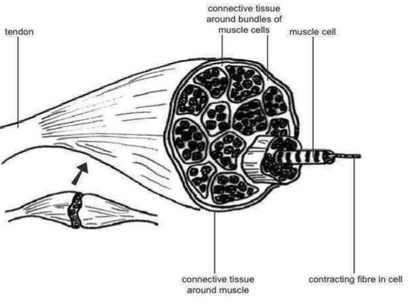
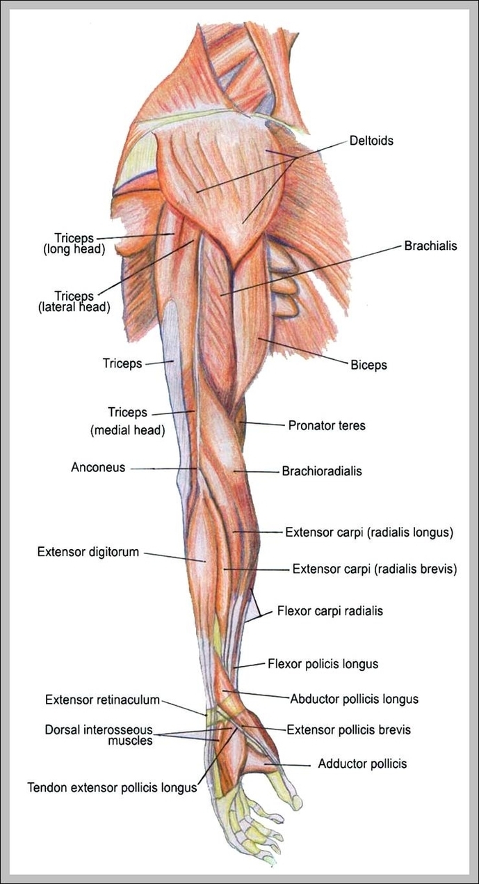
Picture Of Arm Muscles Image
Picture Of Arm Muscles Image Diagram - Chart - diagrams and charts with labels. This diagram depicts Picture Of Arm Muscles Image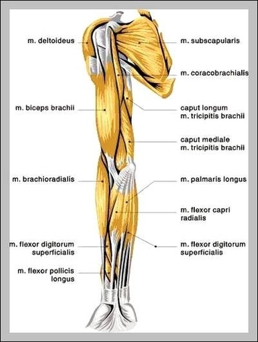
Lower Extremity Muscles Diagram Image
The iliopsoas, an anterior muscle, flexes the thigh. The muscles in the medial compartment adduct the thigh. The illustration below shows some of the muscles of the lower extremity. Muscles that move the leg are located in the thigh region. The quadriceps femoris muscle group straightens the leg at the knee.
When a medical professional refers to your lower extremity, they’re typically referring to everything between your hip to your toes. You lower extremity is a combination of parts: There are over 30 bones in each of your lower extremities including: The muscles in your lower extremity contract and relax to move skeletal bones and thus the body.
The lower leg has two muscle groups to move the ankle, foot, and toes. The bones of the lower leg are the tibia and fibula. The tibia bears most of the weight, and the fibula serves as attachment points for the lower leg muscles. The two tibialis and three peroneus muscles are also found in the lower leg
Lower Extremity Muscles Diagram Image Diagram - Chart - diagrams and charts with labels. This diagram depicts Lower Extremity Muscles Diagram Image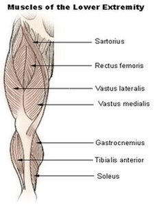
Muscles System1 Image
33,602 muscular system stock photos, vectors, and illustrations are available royalty-free. See muscular system stock video clips
© QA International, 2010. Human muscle system, the muscles of the human body that work the skeletal system, that are under voluntary control, and that are concerned with movement, posture, and balance.
Human muscle system, the muscles of the human body that work the skeletal system, that are under voluntary control, and that are concerned with movement, posture, and balance. Broadly considered, human muscle—like the muscles of all vertebrates—is often divided into striated muscle (or skeletal muscle), smooth muscle, and cardiac muscle.
Muscles System1 Image Diagram - Chart - diagrams and charts with labels. This diagram depicts Muscles System1 Image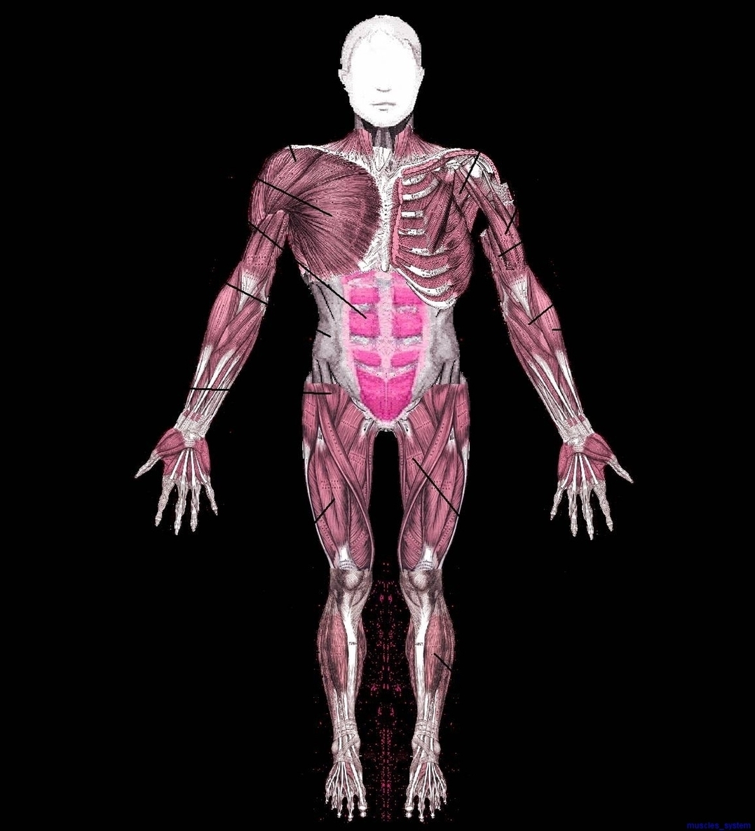
Diagram Back Muscles Image
25,649 back muscle anatomy stock photos, vectors, and illustrations are available royalty-free.
The back supports the weight of the body, allowing for flexible movement while protecting vital organs and nerve structures. This article looks at the anatomy of the back, including bones, muscles, and nerves. It also covers some common conditions and injuries that can affect the back.
There are three different muscle groups found in the back: the superficial group, the deep group, and the intermediate group. Muscles found in the superficial group include rhomboid major, rhomboid minor, levator scapulae, trapezius, latissimus dorsi.
Diagram Back Muscles Image Diagram - Chart - diagrams and charts with labels. This diagram depicts Diagram Back Muscles Image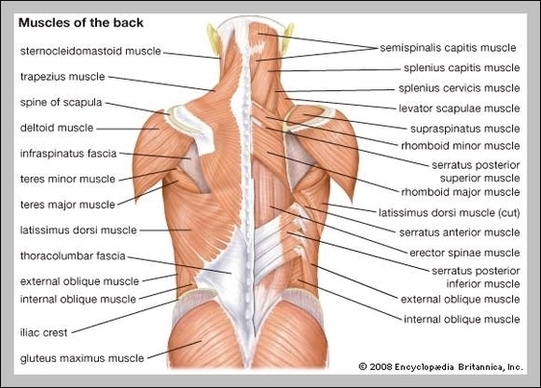
Muscles In Shoulders Image
Muscles In Shoulders Image Diagram - Chart - diagrams and charts with labels. This diagram depicts Muscles In Shoulders Image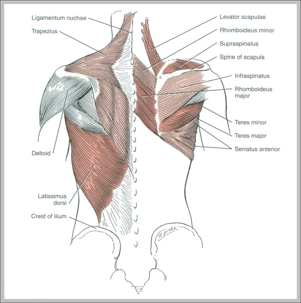
Muscle Tissue Diagram Image
Muscular tissue is a specialized tissue in animals which applies forces to different parts of the body by contraction. It is made up of thin and elongated cells called muscle fibers.
We collected 38+ Muscle Tissue Drawing paintings in our online museum of paintings – PaintingValley.com. A Labelled Diagram O… Draw A Well Labelled…
Last Updated: Jul 16, 2019 The muscular system is responsible for the movement of the human body. Attached to the bones of the skeletal system are about 700 named muscles that make up roughly half of a person’s body weight. Each of these muscles is a discrete organ constructed of skeletal muscle tissue, blood vessels, tendons, and nerves.
Muscle Tissue Diagram Image Diagram - Chart - diagrams and charts with labels. This diagram depicts Muscle Tissue Diagram Image
Skeletal Muscle Diagram Image
Best viewed on 1280 x 768 px resolution in any modern browser. This post is about Skeletal muscle diagram … All diagrams are adhering to our Image Copyright policy.
The skeletal muscle fibers are elongated, cylindrical and multinucleated cells whose length may vary in different animals. In this short guide, you will get a basic concept of skeletal muscle histology from the real slide and labeled diagram.
Skeletal muscles maintain posture, stabilize bones and joints, control internal movement, and generate heat. Skeletal muscle fibers are long, multinucleated cells.
Skeletal Muscle Diagram Image Diagram - Chart - diagrams and charts with labels. This diagram depicts Skeletal Muscle Diagram Image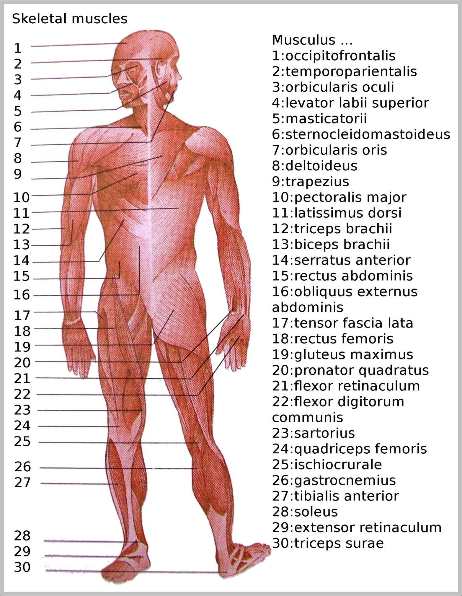
Body Muscles Diagram Image
128,412 muscle anatomy stock photos, vectors, and illustrations are available royalty-free.
5,814 male human anatomy diagram stock photos and images available, or start a new search to explore more stock photos and images. Male silhouette (contour) on white background, vector. Male, female and children’s silhouette on white background,…
Last Updated: Jul 16, 2019 The muscular system is responsible for the movement of the human body. Attached to the bones of the skeletal system are about 700 named muscles that make up roughly half of a person’s body weight. Each of these muscles is a discrete organ constructed of skeletal muscle tissue, blood vessels, tendons, and nerves.
Body Muscles Diagram Image Diagram - Chart - diagrams and charts with labels. This diagram depicts Body Muscles Diagram Image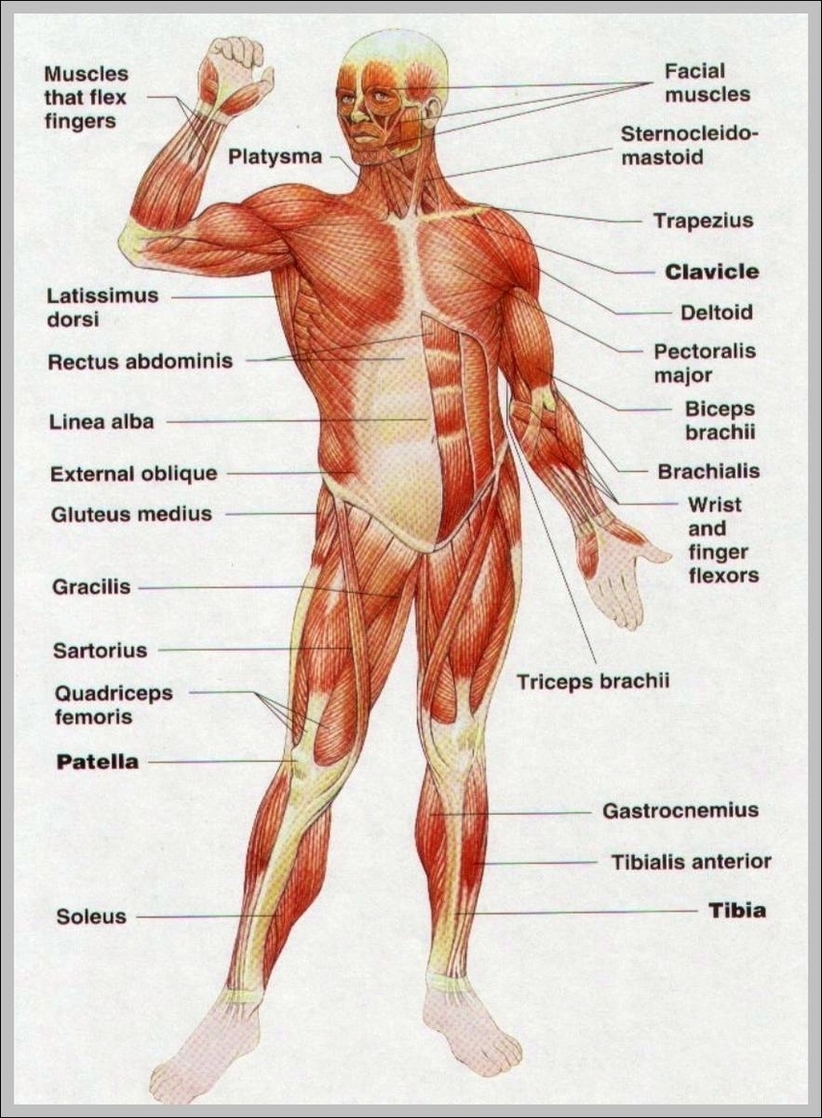
Anatomy Of Leg Muscles Image
Anatomy Of Leg Muscles Image Diagram - Chart - diagrams and charts with labels. This diagram depicts Anatomy Of Leg Muscles Image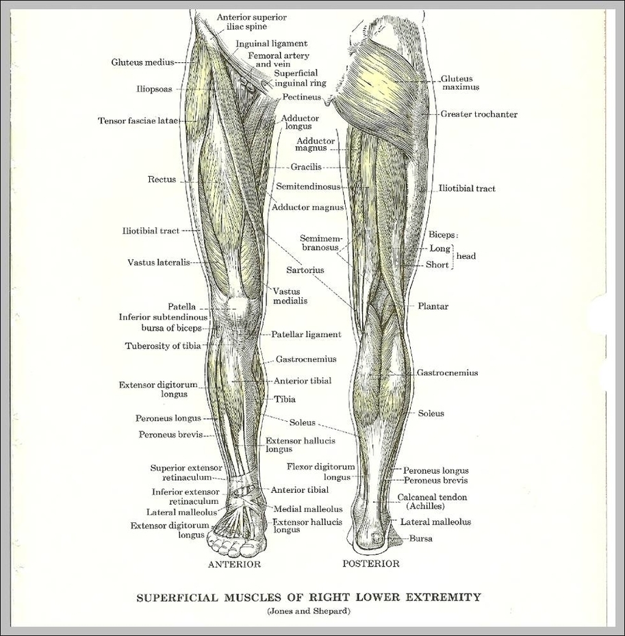
Pull Muscles Image
A pulled muscle occurs when a muscle anywhere in the body is stretched beyond its means, leading to slight tearing of the tiny fibers that make up the muscle. A pulled muscle is sometimes known as a strained muscle, and the condition can be quite painful, though some instances of a pulled muscle will result in very little pain.
Pull ups are a compound movement As you can see above, the exercise incorporates multiple muscle groups at one time, opening itself up for the many benefits of compound movements such as: Increased muscle growth – as all of the above muscles are getting a workout at once, rather than isolation exercises targeting one muscle at a time.
What muscles do pull ups work? 1 Lats (latissimus dorsi) 2 Traps (Trapezius) 3 Rhomboids (Rhomboideus major and minor) 4 Posterior deltoid 5 Biceps (Biceps brachii) 6 Teres Major More …
Pull Muscles Image Diagram - Chart - diagrams and charts with labels. This diagram depicts Pull Muscles Image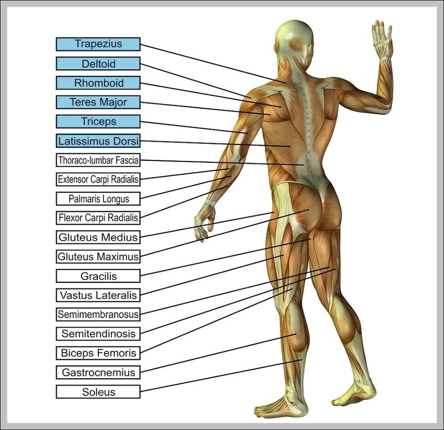
Human Muscles Image
93,451 human body muscles stock photos and images available, or search for anatomy or human anatomy to find more great stock photos and pictures.
Last Updated: Jul 16, 2019 The muscular system is responsible for the movement of the human body. Attached to the bones of the skeletal system are about 700 named muscles that make up roughly half of a person’s body weight. Each of these muscles is a discrete organ constructed of skeletal muscle tissue, blood vessels, tendons, and nerves.
Muscles are the only tissue in the body that has the ability to contract and therefore move the other parts of the body. Related to the function of movement is the muscular system’s second function: the maintenance of posture and body position. Muscles often contract to hold the body still or in a particular position rather than to cause …
Human Muscles Image Diagram - Chart - diagrams and charts with labels. This diagram depicts Human Muscles Image
Female Muscle Anatomy Diagram Image
Female Muscle Anatomy Diagram Image Diagram - Chart - diagrams and charts with labels. This diagram depicts Female Muscle Anatomy Diagram Image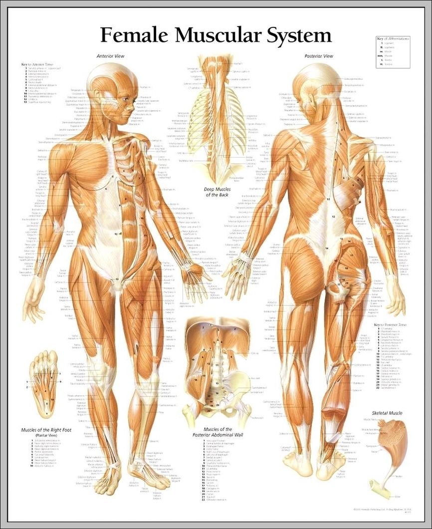
Arms Muscles Image
27,539 arm muscle anatomy stock photos, vectors, and illustrations are available royalty-free.
What Are the Muscles of Your Arms? There are four main muscles of your arms: biceps, triceps, forearm flexors, and forearm extensors. There are also a handful of other muscles that support these main four.
Anterior arm muscles The body’s anterior muscles tend to be the flexors — they pull your extremities inward, toward your center. So the biceps of the upper arms flex (bend) the elbow, and the forearm flexors on the inside of your forearms flex the wrist and fingers.
Arms Muscles Image Diagram - Chart - diagrams and charts with labels. This diagram depicts Arms Muscles Image