Tooth Anatomy

This human anatomy diagram with labels depicts and explains the details and or parts of the Tooth Anatomy. Human anatomy diagrams and charts show internal organs, body systems, cells, conditions, sickness and symptoms information and/or tips to ensure one lives in good health.

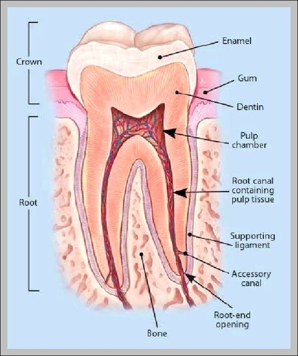
Each part of a tooth has unique functions and properties. Aetna’s Simple Steps to Better Dental Health lists major parts of tooth anatomy, including enamel, dentin, cementum, root (s) and the root canal chamber (s) inside the tooth. Damaged teeth, especially teeth with cracked or eroded enamel, are very susceptible to cavities.

Dental anatomy. Usually, there are 20 primary (“baby”) teeth and 28 to 32 permanent teeth, the last four being third molars or ” wisdom teeth “, each of which may or may not grow in. Among primary teeth, 10 usually are found in the maxilla (upper jaw) and the other 10 in the mandible (lower jaw).

The periodontium consists of: Gingiva: it is a part of the oral mucosa that surrounds teeth and covers the maxilla and mandible. The beautiful smile comes from a harmonious coexistence of teeth and gingiva. Cementum: it is a thin layer that covers the roots of the tooth. Also, it attaches the tooth to the bone.

Tooth Anatomy

tooth anatomy


Tags: , , , , , ,