Thyroid Gland Scan Image

This human anatomy diagram with labels depicts and explains the details and or parts of the Thyroid Gland Scan Image. Human anatomy diagrams and charts show internal organs, body systems, cells, conditions, sickness and symptoms information and/or tips to ensure one lives in good health.

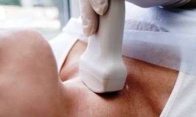
An ultrasound is a painless procedure that uses sound waves to generate images of the inside of your body. Your doctor will often use an ultrasound to create images of a fetus during pregnancy. A thyroid ultrasound is used to examine the thyroid for abnormalities, including:

A doctor who specializes in nuclear imaging will evaluate the images and results of your thyroid scan. Your results will be sent in a report to your doctor. A normal thyroid scan would show no abnormalities in the size, shape, and location of the thyroid gland. Your thyroid will have an even green color on the image.

Ultrasound normal thyroid gland has an homogenous appearance, the capsule may appear as a thin hyperechoic line each lobe normally measures 4 length: 4-7 cm volume (excluding isthmus, unless its thickness is >3 mm) 6 10-15 mL for females

Thyroid Gland Scan Image

Thyroid Gland Scan Image


Tags: , ,