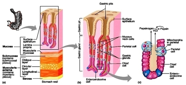
Stomach histology. The stomach is a key part of the gastrointestinal (GI) tract, sitting between the esophagus and duodenum. Its functions are to mix food with stomach acid and break food down into smaller particles using chemical and mechanical digestion. The stomach can perform these roles due to the layers of the stomach wall.
These are the gastric mucosa, submucosa, muscularis externa and serosa. All parts of the GI tract tend to follow this same pattern of tissue layer arrangement, which means that the stomach is essentially just a widening of the GI tube. These layers are best observed when you’re looking at the microanatomy, or histology, of the stomach.
This layered arrangement follows the same general structure in all regions of the stomach, and throughout the entire gastrointestinal tract. The outer layer of the stomach wall is smooth, continuous with the parietal peritoneum.
Stomach Histology Diagram Image

