The Human Eye And Internal Anatomy Of The Eyeball Images Image

This human anatomy diagram with labels depicts and explains the details and or parts of the The Human Eye And Internal Anatomy Of The Eyeball Images Image. Human anatomy diagrams and charts show internal organs, body systems, cells, conditions, sickness and symptoms information and/or tips to ensure one lives in good health.

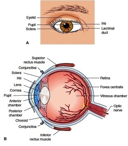
Picture of Eye Anatomy Detail The eye is our organ of sight. The eye has a number of components which include but are not limited to the cornea, iris, pupil, lens, retina, macula, optic nerve, choroid and vitreous. Cornea: clear front window of the eye that transmits and focuses light into the eye.

The Human Eye (Eyeball) Diagram, Parts and Pictures. The human eye consists of the eyeball, optic nerve, orbit and appendages (eyelids, extraocular muscles and lacrimal glands). While the eyeball is the actual sensory organ, the other parts of of the eye are equally important in maintaining the health and function of the eye as a whole.

While the eyeball is the actual sensory organ, the other parts of of the eye are equally important in maintaining the health and function of the eye as a whole. The structure of the human eye is such that light can enter, be refracted and trigger nerve impulses back to the brain which are then deciphered as images.

The Human Eye And Internal Anatomy Of The Eyeball Images Image

The Human Eye And Internal Anatomy Of The Eyeball Images Image


Tags: , , ,