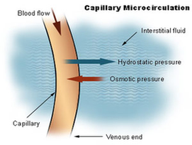
Scott Sundick, MD, is a board-certified vascular and endovascular surgeon. He currently practices in Westfield, New Jersey. Capillaries are the smallest blood vessels in the body, connecting the smallest arteries to the smallest veins. These vessels are often referred to as the “microcirculation.”
Microcirculation Structure and Function. The microcirculation is comprised of arterioles, capillaries, venules, and terminal lymphatic vessels. Small precapillary resistance vessels (10-200 μ) composed of an endothelium surrounded by one or more layers of smooth muscle cells.
They are present in muscle, skin, fat, and nerve tissue. Fenestrated: These capillaries have small pores that allow small molecules through and are located in the intestines, kidneys, and endocrine glands. Sinusoidal or discontinuous: These capillaries have large open pores—large enough to allow a blood cell through.
Capillary Microcirculation Diagram Image

