Scalenes Muscles Image

This human anatomy diagram with labels depicts and explains the details and or parts of the Scalenes Muscles Image. Human anatomy diagrams and charts show internal organs, body systems, cells, conditions, sickness and symptoms information and/or tips to ensure one lives in good health.

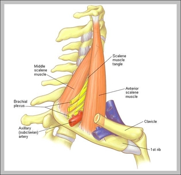
Description. Scalene are a group of three pairs of muscles in the lateral neck: scalenus anterior, scalenus medius and scalenus posterior. Sometimes a fourth muscle, the scalenus minimus is present behind the lower portion of the scalenus anterior. The brachial plexus and subclavian artery pass between the anterior and middle scalenes,…

The Three Scalene Muscles 1 Anterior Scalenes. Of the three muscles that make up the scalenes, the anterior is located closest to the front. … 2 Medial Scalenes. The medial scalene (scalenus medius) resides between the anterior and posterior branches of the scalene muscle. 3 Posterior Scalenes. … 4 The Scalene Triangle. …

Like all scalene muscles, it is considered a side neck muscle. It has more than one function. It’s helpful to remember that you have these muscles on both sides of your neck. When acting only on one side of your neck, the anterior scalene can bend and rotate your neck. When both of your anterior scalenes work together, they flex (bend) your neck.

Scalenes Muscles Image

Scalenes Muscles Image


Tags: ,