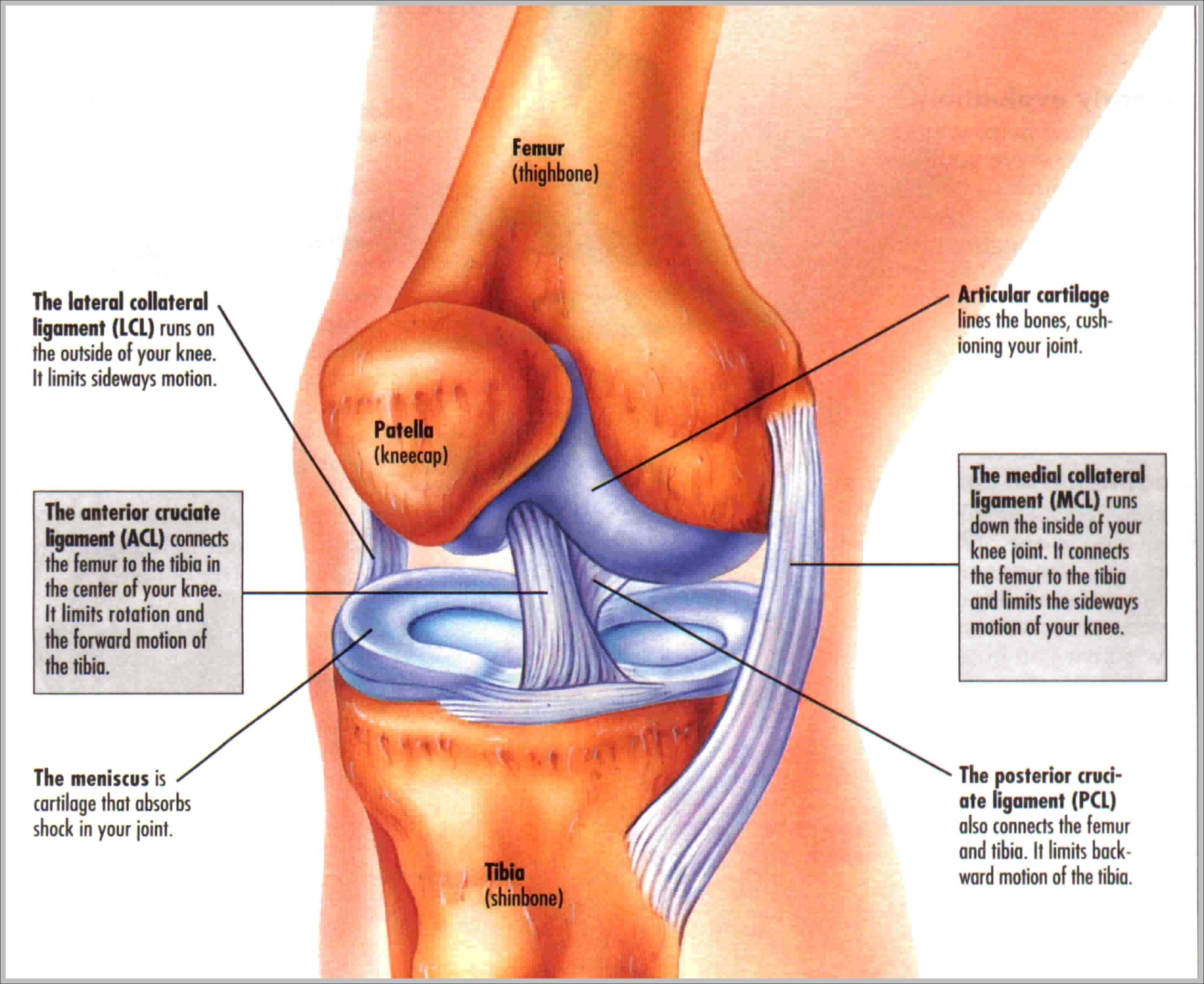
Knee joint: The knee joint has three parts. The thigh bone (the femur) meets the large shin bone (the tibia) to form the main knee joint. This joint has an inner (medial) and an outer (lateral) compartment. The kneecap (the patella) joins the femur to form a third joint, called the patellofemoral joint.
Picture of Knee Joint. The thigh bone (the femur) meets the large shin bone (the tibia) to form the main knee joint. This joint has an inner (medial) and an outer (lateral) compartment. The kneecap (the patella) joins the femur to form a third joint, called the patellofemoral joint. The patella protects the front of the knee joint.
The patella (kneecap) sits over the front of the knee joint. Four major ligaments connect the bones and stabilize the knee joint . In this image, the physician is pointing to the anterior cruciate ligament, or ACL, one of these important ligaments. Inside the knee joint is a smooth cover on the ends of the bone called articular cartilage.
Picture Of Knee

