Bony Landmarks of the Ilium Wing ASIS PSIS Hip Bone.
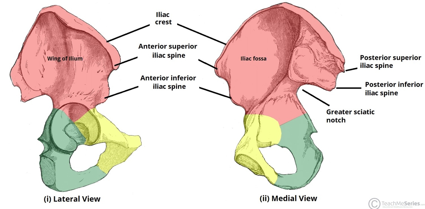
The ilium contains several key bony landmarks that help define the hip bone and provide attachment sites for muscles and ligaments. The wing, or ala, of the ilium forms the broad, flared superior portion and supports the iliac fossa on View Diagram Bony Landmarks of the Ilium Wing ASIS PSIS Hip Bone.