Microscopic Blood Cells

This human anatomy diagram with labels depicts and explains the details and or parts of the Microscopic Blood Cells. Human anatomy diagrams and charts show internal organs, body systems, cells, conditions, sickness and symptoms information and/or tips to ensure one lives in good health.

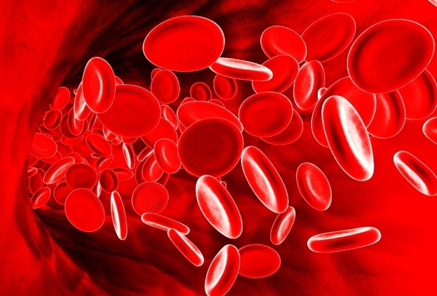
In addition to red blood cells (RBCs), white blood cells (WBCs) and plasma, blood microscopy is believed to show items within the plasma such as: Undigested food particles Fungus Crystals Microbes Bacteria

Look at red blood cells under a microscope Preparation and staining of blood smear. There are over 5 million red blood cells per cubic millimeter of blood. If you just add a drop of blood on a microscopic slide, the specimen will be too “thick” (too many cells) to observe. For this reason, making a diluted specimen is generally required.

The blood cells are mainly red blood cells (also called RBCs or erythrocytes), white blood cells (also called WBCs or leukocytes), and platelets. The red blood cells constitute about 45% of whole blood volume, the plasma about 54%, and white cells about 1%.

Microscopic Blood Cells

Red Blood Cell Image


Tags: