Intermediate Heart Development Timeline Image

This human anatomy diagram with labels depicts and explains the details and or parts of the Intermediate Heart Development Timeline Image. Human anatomy diagrams and charts show internal organs, body systems, cells, conditions, sickness and symptoms information and/or tips to ensure one lives in good health.

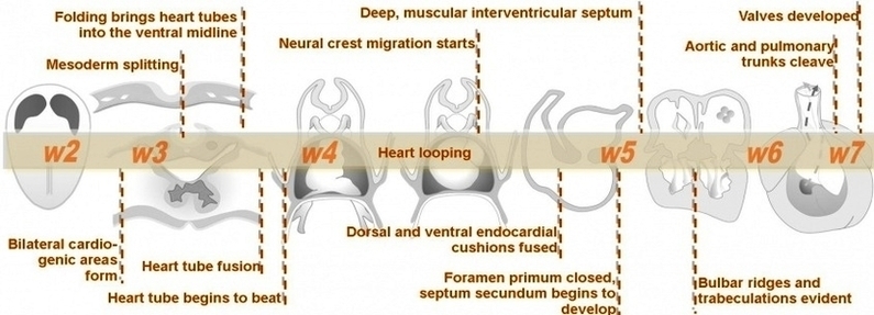
Heart development (also known as cardiogenesis) refers to the prenatal development of the heart. This begins with the formation of two endocardial tubes which merge to form the tubular heart, also called the primitive heart tube.

Endocardial tubes. This early development is critical for subsequent embryonic and prenatal development. The heart is the first functional organ to develop and starts to beat and pump blood at around day 21 or 22. The heart begins to develop near the head of the embryo in the cardiogenic area.

The critical early development of the heart is reflected by the prominent heart bulge that appears on the anterior surface of the embryo. The heart forms from an embryonic tissue called mesoderm around 18 to 19 days after fertilization.

Intermediate Heart Development Timeline Image

Intermediate Heart Development Timeline Image


Tags: , , ,