Diagram Of Heel Pain Exercise Image

This human anatomy diagram with labels depicts and explains the details and or parts of the Diagram Of Heel Pain Exercise Image. Human anatomy diagrams and charts show internal organs, body systems, cells, conditions, sickness and symptoms information and/or tips to ensure one lives in good health.

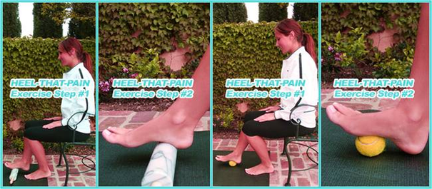
If you are suffering from pain in both heels, you can do this stretch on both feet. This exercise slowly stretches the plantar fascia ligament. Additionally, if you freeze the water bottle prior to doing this heel pain stretch, you can ice your foot while you exercise to minimize the pain and inflammation that is common in Plantar Fasciitis.

This heel pain exercise gently stretches the plantar fascia ligaments, the Achilles’ tendon and the calf muscles, promoting better elasticity and healing. The Wall Stretch also has the added bonus of helping you build new, strong muscle tissue in your calves. Take a seat in a comfortable chair.

A foot pain diagram is a great tool to help you work out what is causing your ankle and foot pain. There are a whole range of structures e.g. bones, muscles, tendons and nerves which will each give slightly different foot pain symptoms.

Diagram Of Heel Pain Exercise Image

Diagram Of Heel Pain Exercise Image


Tags: , ,