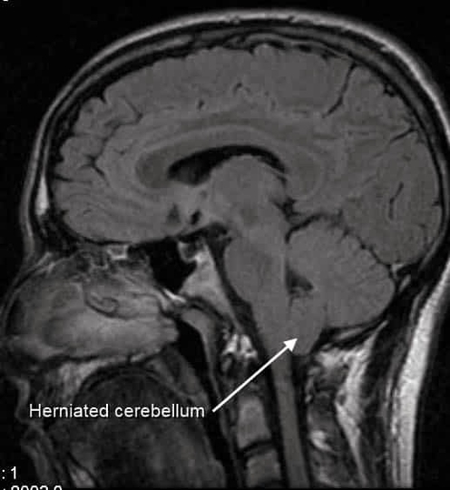
CT scans of cerebellar tonsil herniation show downward displacement through the foramen magnum, often associated with Chiari malformations. This can compress the brainstem, cerebellum, and cerebrospinal fluid pathways, causing headache, dizziness, motor or sensory deficits, and hydrocephalus. Imaging demonstrates the extent of herniation, associated syringomyelia, or crowding of posterior fossa structures. Precise anatomical knowledge of the cerebellum, foramen magnum, and surrounding vessels is crucial for neurosurgeons performing decompression procedures. Early detection allows timely intervention to relieve pressure, restore CSF flow, and preserve neurological function, while avoiding complications such as brainstem injury or persistent neurological deficits. CT Scan of Cerebellar Tonsil Herniation Diagram - Chart - diagrams and charts with labels. This diagram depicts CT Scan of Cerebellar Tonsil Herniation and explains the details of CT Scan of Cerebellar Tonsil Herniation.
CT Scan of Cerebellar Tonsil Herniation