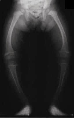
X-rays of rickets reveal characteristic changes in bones due to vitamin D deficiency, including widened, cupped, and frayed metaphyses, especially at the wrist and knee. Rickets affects bone mineralization in children, leading to deformities like bowing of long bones and delayed growth. Knowledge of radiographic features is essential for pediatricians, orthopedic surgeons, and medical students in diagnosing rickets, planning treatment with vitamin D and calcium supplementation, and monitoring skeletal development. Recognizing these X-ray patterns ensures timely intervention, prevents long-term deformities, and guides assessment of treatment efficacy in affected children. X Ray of Rickets Diagram - Chart - diagrams and charts with labels. This diagram depicts X Ray of Rickets and explains the details of X Ray of Rickets.
X Ray of Rickets