Connective Tissue2 Diagram Image

This human anatomy diagram with labels depicts and explains the details and or parts of the Connective Tissue2 Diagram Image. Human anatomy diagrams and charts show internal organs, body systems, cells, conditions, sickness and symptoms information and/or tips to ensure one lives in good health.

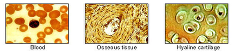
20,642 connective tissue stock photos and images available, or search for connective tissue disease or connective tissue massage to find more great stock photos and pictures. Areolar connective tissue under the microscope view.

They are loose connective tissues and dense or fibrous connective tissues. There are three types of loose connective tissues and these are areolar connective tissues, adipose connective tissues, and reticular tissues. Areolar connective tissue – The areolar connective tissue is a loose array of fibers consists of various types of cells.

This image of loose connective tissue shows collagenous fibers (red), elastic fibers (black), matrix, and fibroblasts (cells that produce the fibers). Ed Reschke/Photolibrary/Getty Images In vertebrates, the most common type of connective tissue is loose connective tissue.

Connective Tissue2 Diagram Image

Connective Tissue2 Diagram Image


Tags: ,