
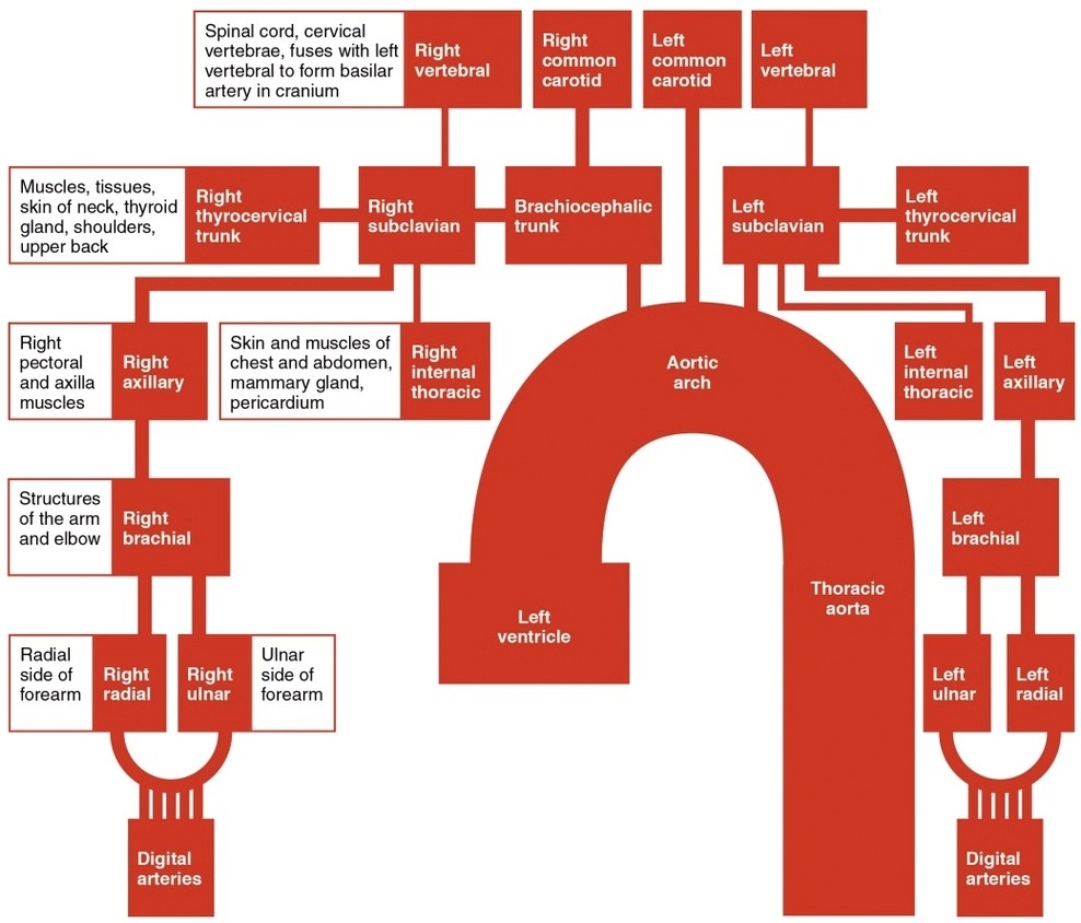
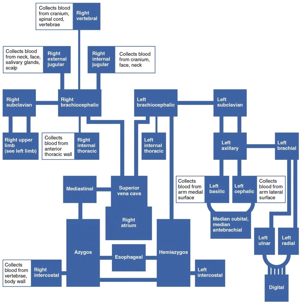

Thoracic Upper Limb Veins
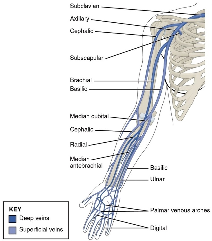
Thoracic Upper Limb Veins: The thoracic and upper limb veins include the subclavian, axillary, brachial, cephalic, and basilic veins, which return blood from the arms and upper chest to the heart via the superior vena cava.