
Category Archives: Anatomy
Dental Restoration Oceanside Ca Image

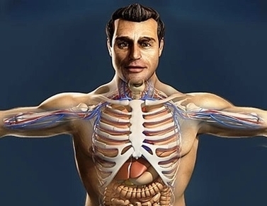
Atlas Of Human Anatomy Image
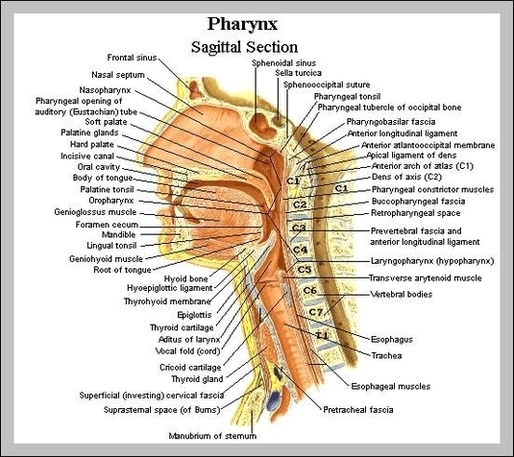
Images in: CT, MRI, Radiographs, Anatomic diagrams and nuclear images. Available in 12 languages. Interactive anatomical atlas of the head, brain, and neck based on anatomical diagrams and CT and MRI medical imaging exams 28,679 anatomy of the human body View Diagram Atlas Of Human Anatomy Image
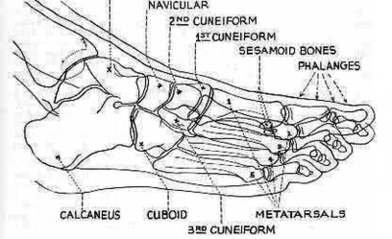
Foot Dpobl Anatomy Image
Embryo Times Life Size Image
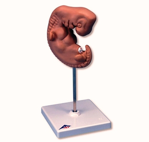
42,318 embryo stock photos, vectors, and illustrations are available royalty-free. See embryo stock video clips The embryo measures about 4 mm (1/8 inch) from head to rump, and is about the size of a grain of rice. Fetus Illustration Embryo View Diagram Embryo Times Life Size Image
Atlas Tone Abdominal Muscles Image
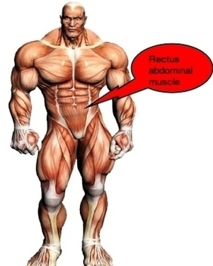
The Atlas Muscle Toner is clinically demonstrated to deliver firmer, stronger and more toned abdominal muscles while you are: at home, at work, watching TV, exercising, folding laundry, helping your kids with their homework, taking a walk…Doing virtually anything! Atlas View Diagram Atlas Tone Abdominal Muscles Image
Ankle Sprain Causes Image
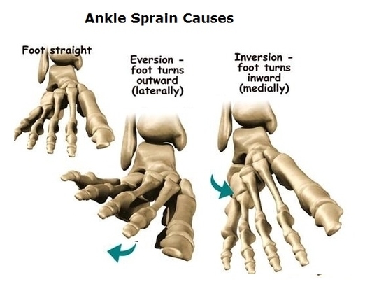
A sprain occurs when your ankle is forced to move out of its normal position, which can cause one or more of the ankle’s ligaments to stretch, partially tear or tear completely. Causes of a sprained ankle might include: Factors View Diagram Ankle Sprain Causes Image
Dap Bottle Image Image

plastic bottle cap images 135,224 plastic bottle cap stock photos, vectors, and illustrations are available royalty-free. See plastic bottle cap stock video clips of 1,353 Get your bottle caps and epoxy at www.fizzypops.com. Rhiley’s Loops is now donating FREE BOTTLE View Diagram Dap Bottle Image Image
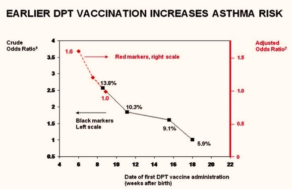
Earlier Vaccination Causes Asthma Diagram Image
Stent Graft Image
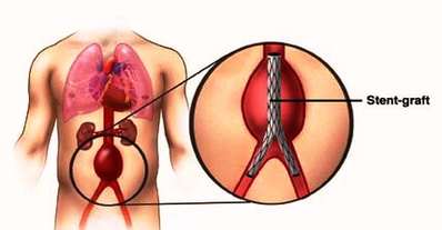
A stent-graft is a device constructed from a stent and a vascular conduit that is inserted using catheter techniques and image guidance. The stent serves to anchor the graft in the blood vessel lumen, and in most cases provides structural View Diagram Stent Graft Image
Anatomy Semi Textured Image
Gum Disease Closeup Image
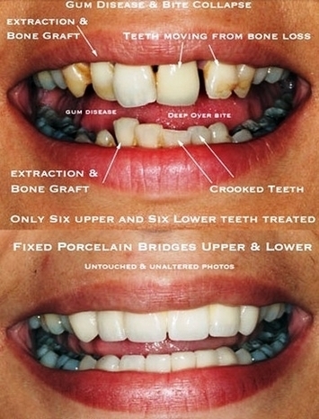
5,270 gum disease stock photos and images available, or search for stages of gum disease or gum disease vector to find more great stock photos and pictures. Gum bleeding and inflammation close up. A man examined by a dentist. The View Diagram Gum Disease Closeup Image
Healthy Habits Chart Photos Image
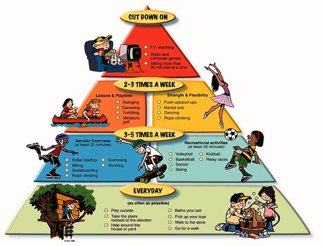
2,749,779 healthy habits stock photos and images available, or search for healthy food or wellness to find more great stock photos and pictures. Healthy lifestyle habits colorful line vector icons isolated. Proper nutrition fruit vegetables water seafood. Physical activity sport View Diagram Healthy Habits Chart Photos Image
Final Long Bone Diagram Image
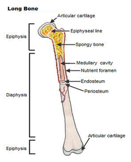
Long bone anatomy A long bone is a bone that has greater length than width. A long bone has a shaft and 2 ends. Long bones have a thick outside layer of compact bone and an inner medullary cavity containing View Diagram Final Long Bone Diagram Image
Steroids To Minimize Asthma Attack Relapses Image
Inhaled steroids are the mainstay treatment for controlling asthma. The use of inhaled steroids leads to: Steroids may help asthma symptoms during an attack but are slow acting and can take several hours to take effect. Dosages of inhaled steroids View Diagram Steroids To Minimize Asthma Attack Relapses Image
Austin Cyberknife Prostate Cancer Staging Image
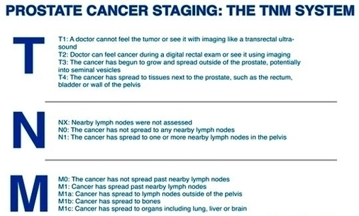
The CyberKnife System is a non-invasive, non-surgical, pain-free treatment option for prostate cancer with proven results. It is the first and only radiotherapy device that brings true robotic precision and the ability to track, detect and correct for tumor motion View Diagram Austin Cyberknife Prostate Cancer Staging Image
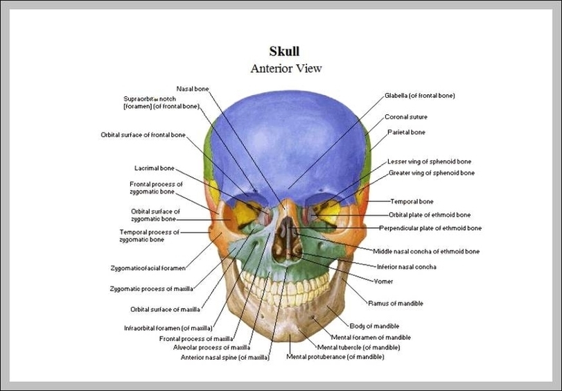
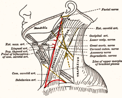
Head And Neck Anatomy Image
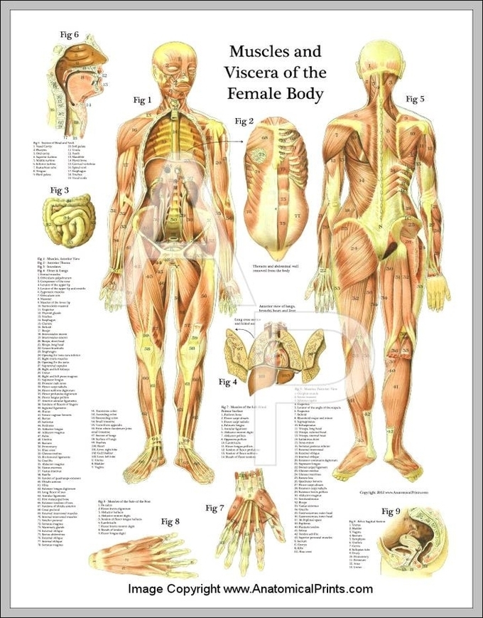
Female Muscle Anatomy Chart Image
Dangers High Blood Pressure Image
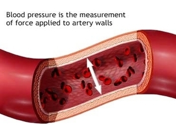
Given that high blood pressure significantly increases the risk for both heart attack and stroke, there’s no question that it’s a serious and often deadly concern. In fact, AHA researchers say that, in 2015, about 79,000 Americans died as a View Diagram Dangers High Blood Pressure Image
Diagram Gyn Symptoms Matrix Image
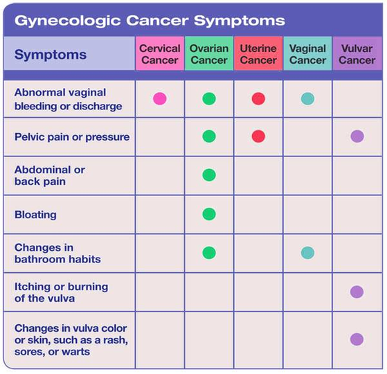
The matrix diagram shows the relationship between two, three, or four groups of information. It also can give information about the relationship, such as its strength, of the roles played by various individuals or measurements. L-shaped matrix diagram What is View Diagram Diagram Gyn Symptoms Matrix Image