
Inner Organs Diagram
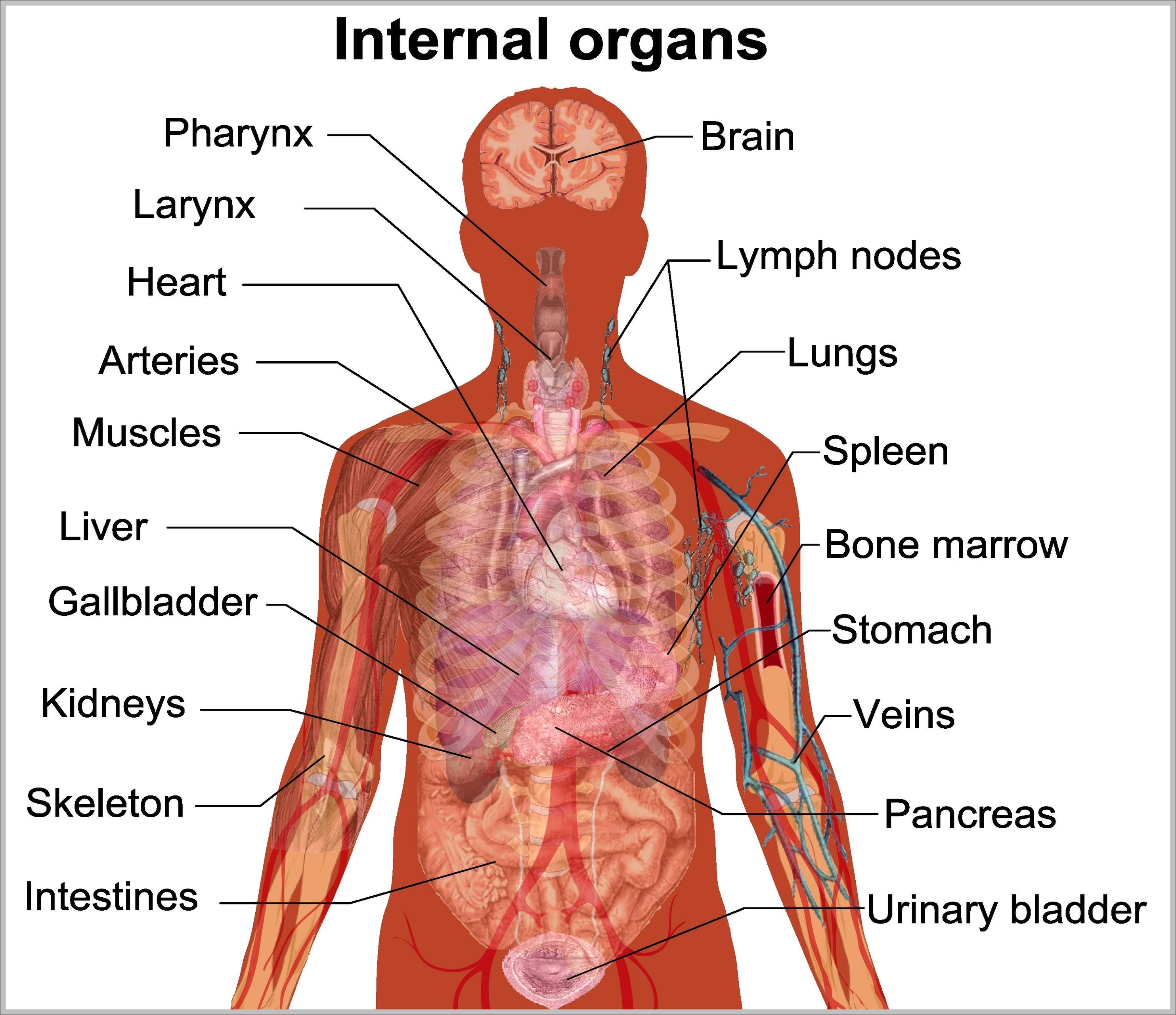
Inner Organs Diagram: An inner organs diagram reveals the internal organs such as the lungs, heart, stomach, intestines, kidneys, and liver arranged within the torso.
Inner Organs Diagram: An inner organs diagram reveals the internal organs such as the lungs, heart, stomach, intestines, kidneys, and liver arranged within the torso.

Inner Ear Anatomy: The inner ear consists of the cochlea, vestibule, and semicircular canals. It is responsible for hearing and balance.
Inner Body Anatomy: The inner body anatomy encompasses all of the internal systems and organs that work together to keep the human body functioning, including the cardiovascular, digestive, respiratory, and nervous systems, among others.
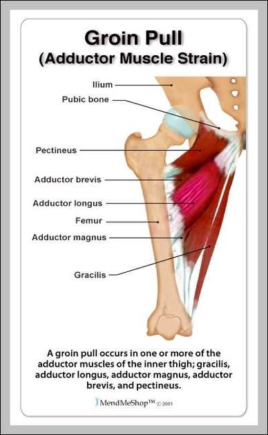
Inguinal Muscle: The inguinal area includes the inguinal ligament and associated muscles like the internal oblique, which support the lower abdominal wall and play a role in core stability.

Inguinal Ligament Exercises: Strengthening exercises target muscles around the inguinal ligament, including hip flexors and core, to support pelvic stability and reduce strain.

Immune System Diagram: An immune system diagram shows the components of the immune system, including white blood cells, lymph nodes, spleen, and antibodies. It illustrates how the body defends against pathogens and maintains health.
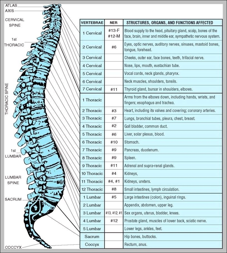
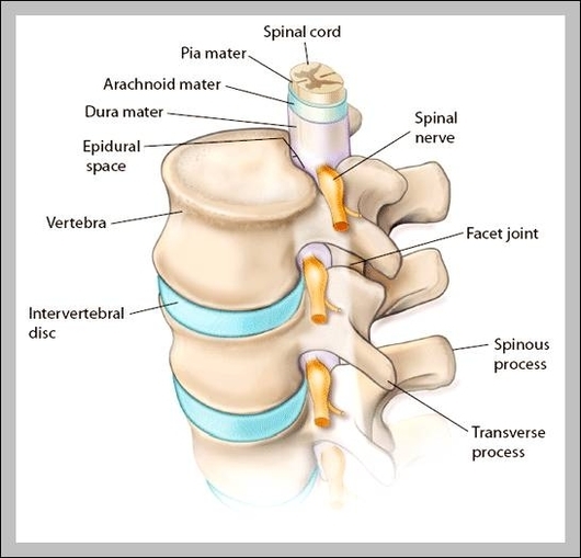
Images Of The Spine: Images of the spine show the vertebrae, discs, and spinal cord. The spine plays a key role in supporting the body, protecting the spinal cord, and enabling movement.
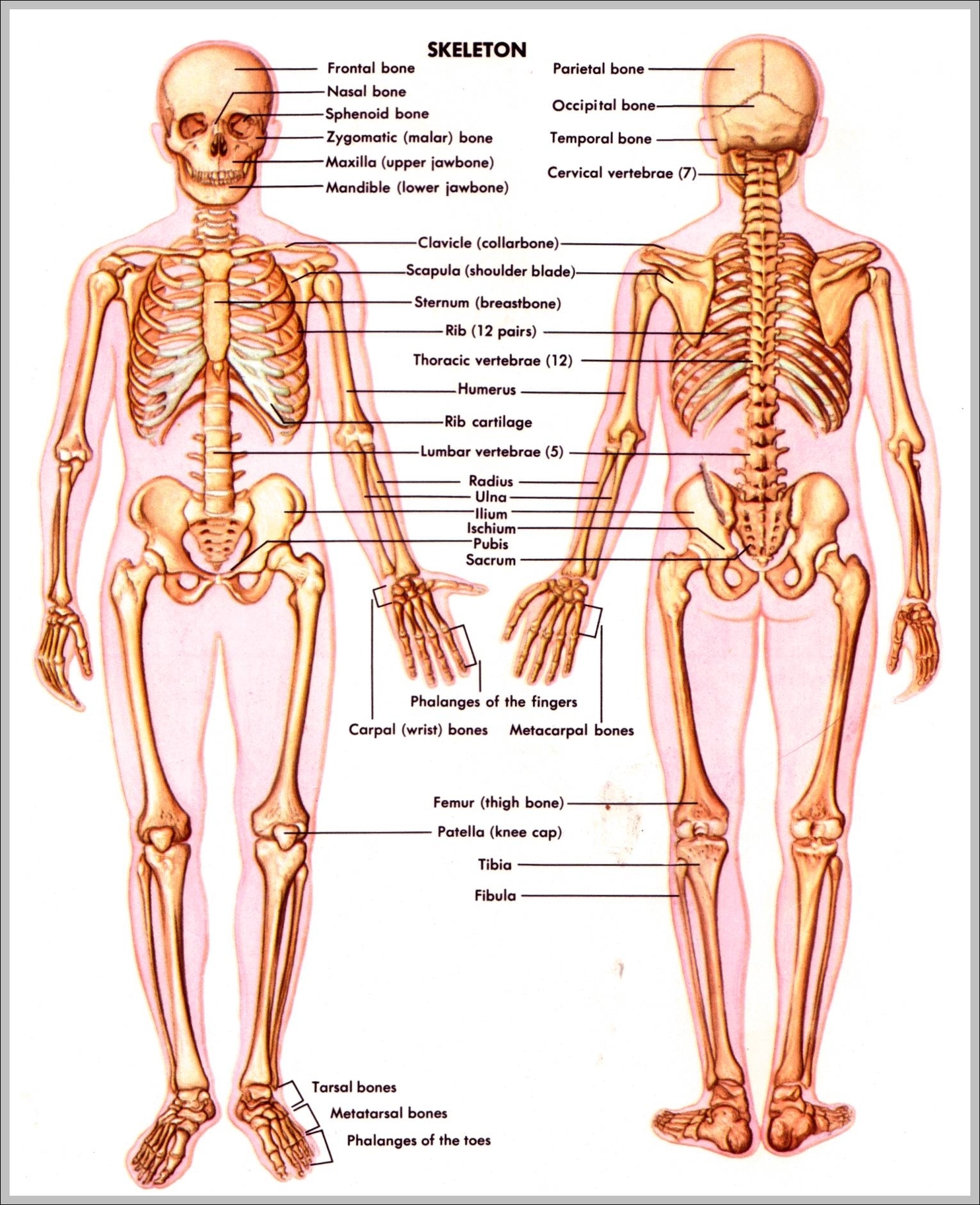
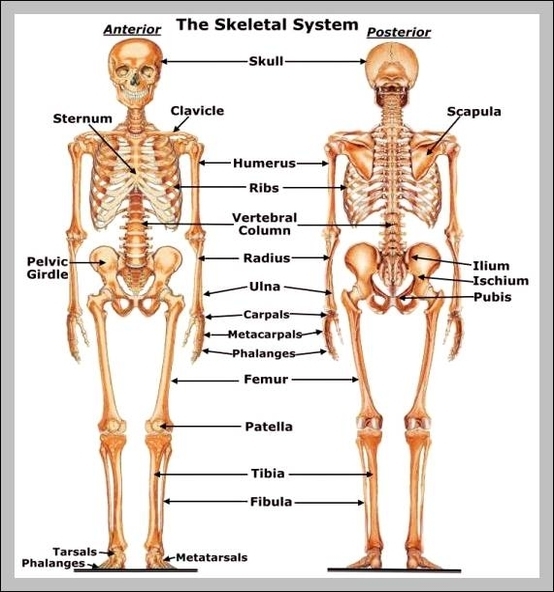
Images Of The Skeletal System: Images of the skeletal system show the bones, joints, and ligaments that make up the bodys framework, providing support and enabling movement.
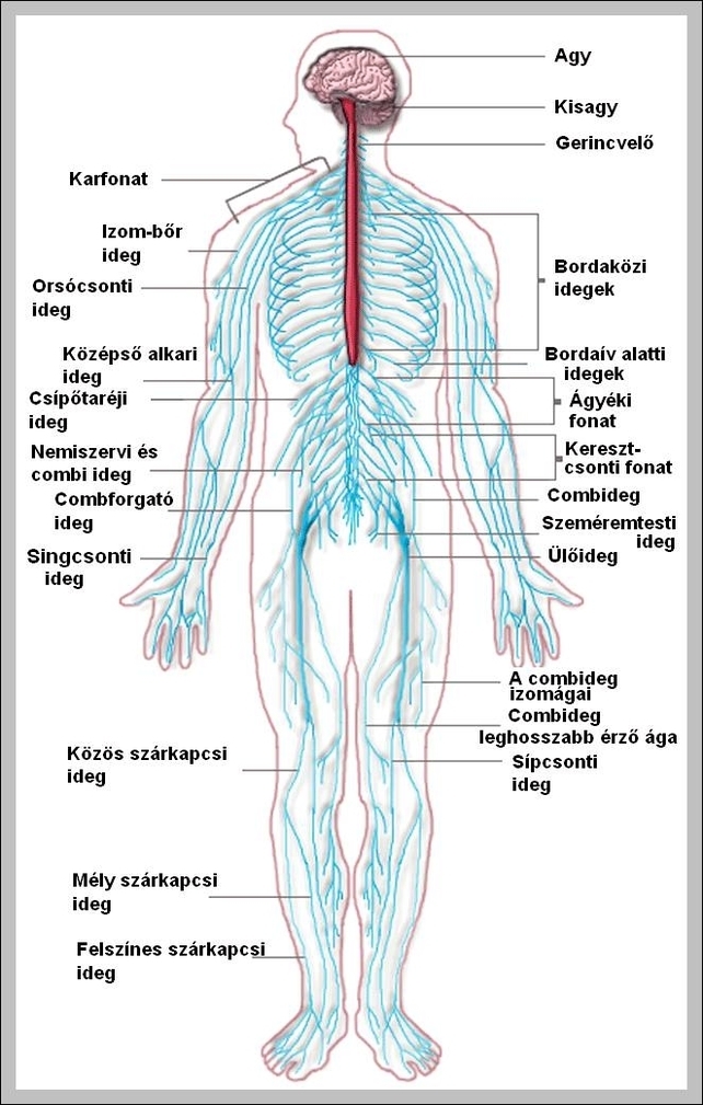
Images Of The Nervous System: These images illustrate brain regions, the spinal cord, cranial nerves, and peripheral nerve pathways throughout the body.
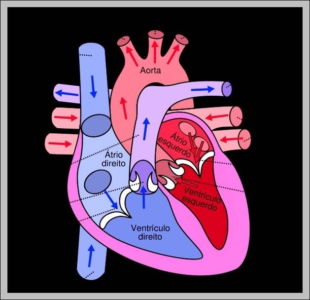
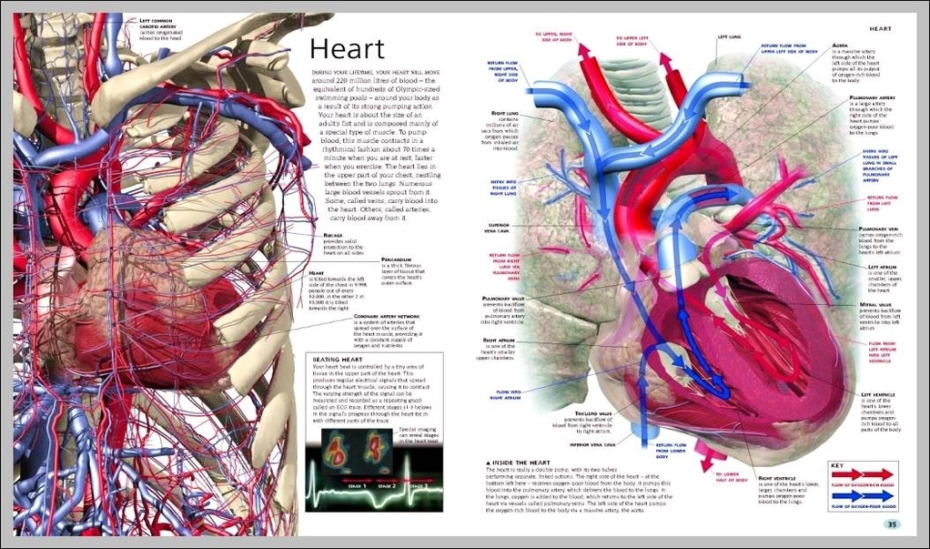
Images Of The Human Heart: Images of the human heart depict its four chambers, valves, and major blood vessels. These visuals are essential for understanding how the heart pumps blood throughout the body and supports cardiovascular health.
Images Of The Human Body For Kids: Images of the human body for kids use simplified visuals and labels to teach basic anatomy such as the head, torso, limbs, and major internal organs.

Images Of The Heart: These images show the heart’s structure, including its four chamberstwo atria and two ventriclesalong with arteries, veins, and valves involved in circulation.

Images Of The Endocrine System: Images of the endocrine system show the glands responsible for hormone production and regulation, such as the thyroid, pituitary, and adrenal glands.
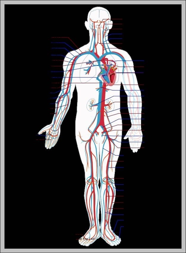
Images Of The Circulatory System: Images of the circulatory system show the heart, arteries, veins, and capillaries, depicting how blood circulates throughout the body to transport oxygen and nutrients.
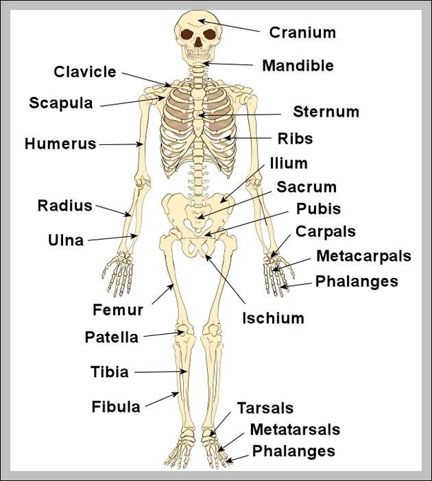
Images Of Skeleton Bones: Images of skeleton bones show the different bones in the human body, from the skull to the extremities, illustrating the structure and arrangement.
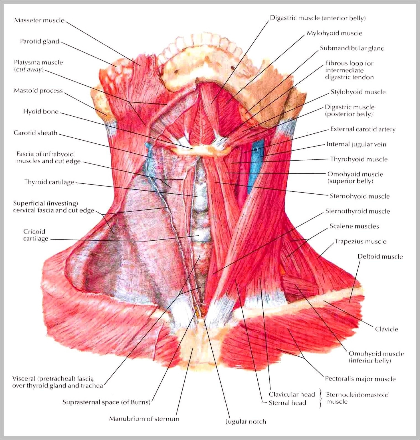
Images Of Neck Muscles: Images of neck muscles show the sternocleidomastoid, trapezius, and scalene muscles, which are important for neck movement and posture.

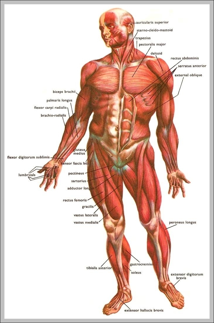
Images Of Muscles: Images of muscles depict the various muscle groups in the body, such as skeletal, smooth, and cardiac muscles, illustrating their structure and function.
Images Of Human Body: Images of the human body provide visual representations of the bodys structure, including bones, muscles, organs, and systems, aiding in the understanding of human anatomy.
Images Of Cervical Spine: Images of the cervical spine highlight the seven vertebrae in the neck region. These vertebrae protect the spinal cord, support the head, and allow for a range of motion, including rotation and flexion of the neck.

Images Of Body: Images of the body may include anatomical drawings, diagrams, or realistic renderings that show body systems, posture, and proportion from different angles.